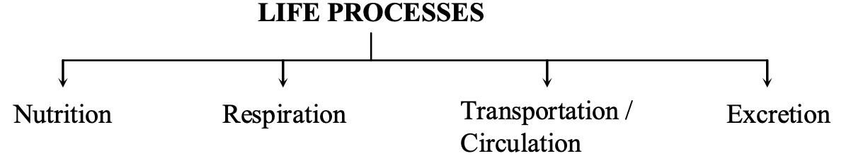
Life Processes: Essential Functions for Survival in Living Organisms
Life Processes form the foundation of Class 10 Science and help students understand how living beings sustain life. The chapter deals with essential biological functions such as nutrition, respiration, transportation, and excretion. Nutrition explains how organisms take in food and utilize it for energy. Plants perform photosynthesis while animals rely on heterotrophic nutrition. Respiration is the process of releasing energy from food, which occurs in two ways: aerobic and anaerobic. This energy is required for every activity in living organisms. The circulatory system explains the transport of nutrients and gases through blood in humans, while in plants, xylem and phloem carry water, minerals, and food. Excretion highlights how living beings remove waste products to maintain balance, including human excretory organs like kidneys, ureters, and bladder, and plants that excrete through diffusion. Understanding life processes is not only essential for board exams but also helps in real-life applications like health and environmental awareness. Students preparing for exams can strengthen their foundation through this chapter and practice diagrams like the human digestive system and nephron structure, which often appear in exams. A quick reference to the NCERT textbook and NCERT solutions for Class 10 Science ensures clarity of concepts and effective revision. Regular study of life processes helps students build confidence for exams and develop a deep understanding of biology.
Understanding Life Processes: The Foundation of Life
Life processes are the essential biological functions that all living organisms perform to maintain their existence on Earth. These fundamental activities distinguish living beings from non-living matter and ensure the continuity of life through proper maintenance and repair of cellular structures.
Living organisms exhibit various characteristics that indicate life, including growth, movement, and metabolism. While visible movement often serves as an obvious sign of life, even seemingly motionless organisms like plants undergo constant molecular movements essential for survival. The maintenance of organized structures requires continuous energy input, obtained through nutrition, to prevent the breakdown of order that would result in death.
The Four Major Life Processes
1. Nutrition: Energy and Building Blocks for Life
Nutrition encompasses the intake and utilization of nutrients by organisms for growth, repair, and energy production. This process involves breaking down complex molecules into simpler, absorbable forms that cells can utilize.
Autotrophic Nutrition occurs in organisms like green plants that synthesize their own food through photosynthesis. Using chlorophyll, plants convert carbon dioxide and water into glucose using sunlight energy. This remarkable process not only feeds the plant but also produces oxygen essential for most life on Earth.
Heterotrophic Nutrition characterizes organisms that obtain food from other sources. This includes various feeding strategies: herbivores consume plants, carnivores feed on other animals, omnivores eat both plants and animals, and saprophytes decompose dead organic matter.
2. Respiration: Cellular Energy Production
Respiration is the catabolic process through which organisms oxidize food molecules to release energy stored as ATP (Adenosine Triphosphate). This process occurs at the cellular level, primarily in mitochondria, often called the "powerhouses of cells."
Aerobic Respiration requires oxygen and completely breaks down glucose into carbon dioxide and water, yielding 38 ATP molecules per glucose molecule. This efficient process powers most cellular activities in complex organisms.
Anaerobic Respiration occurs without oxygen, producing either ethanol (in yeast) or lactic acid (in muscle cells) along with only 2 ATP molecules. While less efficient, this process allows organisms to survive in oxygen-poor environments or during intense physical activity.
3. Transportation: Distribution Throughout the Organism
Transportation systems ensure the distribution of nutrients, gases, and waste products throughout an organism's body.
In Plants, two specialized tissues handle transportation:
- Xylem conducts water and minerals upward from roots through transpiration pull
- Phloem translocates manufactured food (sugars) bidirectionally through the plant
In Animals, particularly humans, the circulatory system comprises:
- Blood vessels (arteries, veins, capillaries) forming an extensive network
- The heart acting as a muscular pump maintaining circulation
- Blood containing plasma, red blood cells (for oxygen transport), white blood cells (for immunity), and platelets (for clotting)
4. Excretion: Waste Removal for Homeostasis
Excretion eliminates harmful metabolic waste products to maintain internal balance. Different organisms employ various strategies:
Plants excrete gaseous wastes through stomata and lenticels, store some wastes in vacuoles, and shed others with falling leaves.
Animals use specialized organs like kidneys (in humans) containing millions of nephrons that filter blood, reabsorb useful substances, and produce urine containing nitrogenous wastes.
Formulas in Life Processes
| Process | Formula | Description |
| Photosynthesis | 6CO₂ + 12H₂O → C₆H₁₂O₆ + 6O₂ + 6H₂O | Conversion of light energy to chemical energy in glucose |
| Aerobic Respiration | C₆H₁₂O₆ + 6O₂ → 6CO₂ + 6H₂O + 38 ATP | Complete oxidation of glucose with oxygen |
| Anaerobic (Yeast) | C₆H₁₂O₆ → 2C₂H₅OH + 2CO₂ + 2 ATP | Fermentation producing ethanol |
| Anaerobic (Muscle) | C₆H₁₂O₆ → 2C₃H₆O₃ + 2 ATP | Lactic acid formation during oxygen debt |
Interdependence of Life Processes
These four fundamental processes work in concert to maintain life. Nutrition provides raw materials, respiration releases energy from these materials, transportation distributes both nutrients and waste products, while excretion removes harmful byproducts. This intricate coordination ensures organisms maintain homeostasis and respond effectively to environmental changes.
Understanding life processes provides crucial insights into how organisms function, adapt, and survive. From the microscopic activities within cells to the complex organ systems of higher organisms, these processes demonstrate the remarkable organization and efficiency of living systems. This knowledge forms the foundation for advances in medicine, agriculture, biotechnology, and environmental conservation.
WHAT ARE LIFE PROCESSES?
If we see a dog running, a bird chirping or a cow chewing cud, we know that these are living beings. How do we know that they are alive, when they are asleep? Yes, we see them breathing. But then, what about plants? They grow over time so they are alive.
In other words, we tend to think of some sort of movement, either growth related or not as a common evidence for being alive. But plants and animals even without visible movement are also alive. So, visible movement as the defining characteristic of life is not enough. Invisible molecular movements are also necessary for life.
Living organisms are well organized structures. But this organized structure is likely to get disturbed over time. If the order breaks down, the organism will no longer be alive. So, living creatures must keep repairing and maintaining their structures.
The various basic functions performed by living organisms to maintain their life on this earth are called life processes. The basic life processes common to all living organisms are as follows:
NUTRITION
"Nutrition" is a process of intake as well as utilization of nutrients by an organism. It is the process of breakdown of nutrients into smaller molecules and their absorption. Food provides us nutrition and energy. It contains different types of nutrients in varying amounts according to the need of our body.
OR
The maintenance processes are needed to prevent damage and break-down for whichenergy in needed. This energy in the form of food comes from outside the body by a process called nutrition.
OR
The process of obtaining food from the surroundings and using it for various metabolic activities by an organism is called nutrition.
Nutrients:
These are the substances required by our body for its growth, repair, work and maintenance of the body. Different types of nutrients are carbohydrates, fats, proteins, vitamins, minerals etc. Our daily energy need may vary according to our occupation, age, sex and under some specific conditions.
MODES OF NUTRITION:
It is defined as the method or mode of obtaining food by an organism. These are of two types.
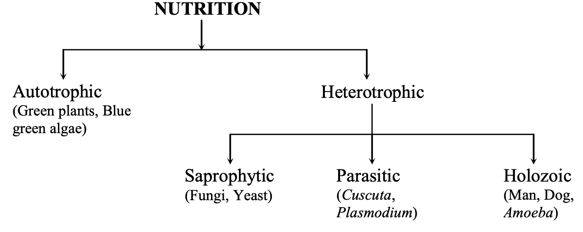
Autotrophic: (Auto = self, trophic = food)
It is a mode of nutrition in which organisms prepare their own food. Inorganic molecules like CO2 and H2O are converted into organic molecules like carbohydrates in the presence of sunlight and chlorophyll e.g. green plants. Autotrophs are further categorized as:
(i) Photoautotrophs: Those which utilize sunlight for preparing their food
(ii) Chemoautotrophs: Those which utilize chemical energy for preparing their food.
Heterotrophic:
(Hetero = different; trophic =food)
It is a mode of nutrition in which organisms derive their food from some other animals or plants. They cannot prepare their own food e.g. human being. Heterotrophs are further categorized depending on the nature of food they consume:
(i) Herbivores: Animals which eat only plants, e.g. cow, goat etc.
(ii) Carnivores: They feed on flesh of other animals, e.g. lion, vulture etc.
(iii) Omnivores: They feed on plants and animals both, e.g. dog, human etc.
(iv) Detritivores: Feed on detritus or dead organic remains, e.g. earthworm etc.
(v) Sanguivorous: Feed on blood, e.g. Leech, female mosquito etc.
(vi) Frugivorous: Feed on fruits, e.g. parrot etc.
(vii) Insectivores: Feed on insects, e.g. bats etc.
On the Basis of Mode of Feeding Organisms are Categorised As:
(a) Saprotrophic Nutrition : It refers to the mode of nutrition in which organisms obtain nutrients from the dead and decaying organic matter e.g. fungi (yeast) and some bacteria. These organisms are called saprophytes. These organisms break-down the food material outside the body and then absorb it.
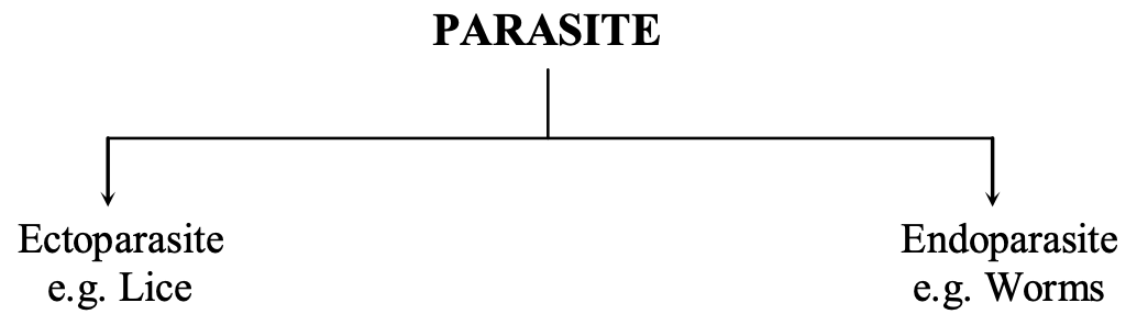
(b) Parasitic Nutrition : It refers to the mode of obtaining food synthesized by other animals and effecting them badly. The organism which obtains food is called the parasite and the organism from which the food is obtained is called ‘host’. This nutrition is observed in fungi, bacteria, few plants like Cuscuta, orchids and some animals like Plasmodium, roundworm, ticks, lick, leeches etc.
Parasite can be further classified as follows
(c) Holozoic Nutrition : it refers to the mode of nutrition in which the complex organic matter in the form of solid food is ingested, digested and then absorbed into the cells and utilized e.g. Amoeba, frog, human being etc. In single-celled organisms, the food may be taken in by the entire surface. But as the complexity of the organism increases different parts become specialized to perform different functions.
Nutrition can be divided into two categories on the basis of occurrence:
(1) Nutrition in plants
(2) Nutrition in animals
NUTRITION IN PLANTS:
- Plants are autotrophic in nature. They prepare their own food hence they are called as producers.
- They contain a green pigment called chlorophyll which can entrap solar energy which is then converted into chemical energy in the form of food and the process is called as "Photosynthesis".
Photosynthesis:
Definition: The synthesis of organic compounds like glucose from simple inorganic molecules like CO2 and H2O by the cells of green plants having chlorophyll in the presence of sunlight is called as photosynthesis.
Equation of Photosynthesis: Photosynthesis is a two step process.
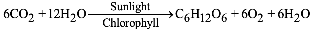
(A) Light reaction: ATP, NADPH2 and O2are produced.
(B) Dark reaction: CO2 & H2O are converted into glucose
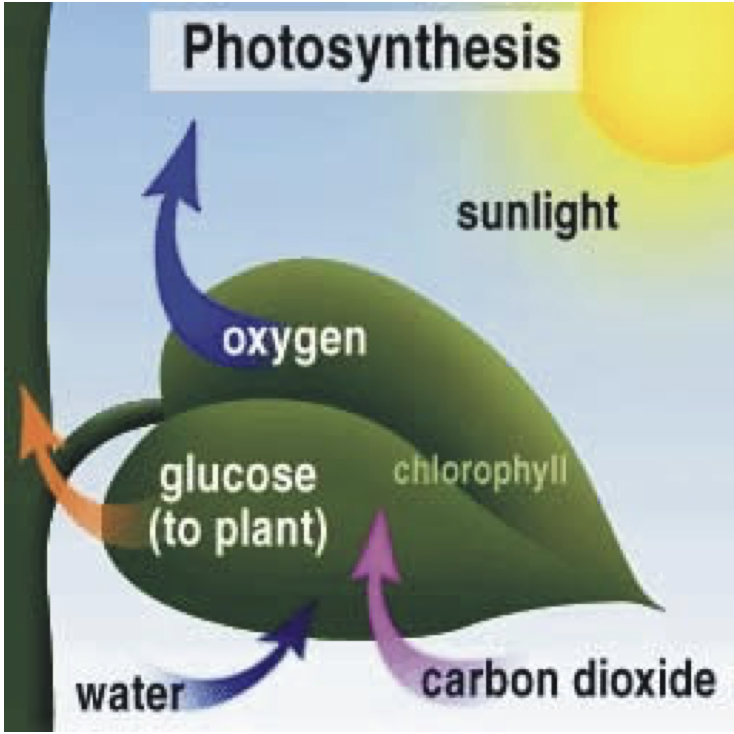
Green plants make their own food by photosynthesis.
(a)Steps of photosynthesis: During the process of photosynthesis, the following events occur :
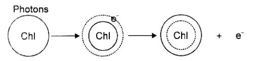
- Absorption of light energy by chlorophyll.
- Conversion of light energy to chemical energy and splitting of water molecules into hydrogen and oxygen.
H2O → 2H+ + 2e– + 1/2 O2
The above processes are considered as light reaction as it can take place only in the presence of light.
- Reduction of carbon-dioxide to carbohydrates: This is also known as dark reaction as it does not require light.
Desert plants take up CO2 at night and prepare an intermediate which is acted upon by the energy absorbed by the chlorophyll during the day.
PHOTOSYNTHESIS ESSENTIALLY REQUIRES TWO THINGS:
Sunlight:
- For plants sun is the basic source of radiant energy.
- Plants utilize the light in the visible region of solar spectra (electromagnetic spectrum) which comes under the range of 400 nm - 700 nm.
- Visible region consists of white light which is a mixture of 7 lights of different wavelengths.
- Maximum photosynthesis occurs in red region.
- There is no photosynthesis in green region because green parts of plants reflect whole of the green light.
Chlorophyll:
These are the green pigments present in chloroplast. They are found in green leaves in a maximum amount as well as in other green aerial parts of plant. There are six different types of chlorophyll, they are chlorophyll a, b, c, d, e and bacteriochlorophyll, amongst them chlorophyll a and chlorophyll b are the most commonly occurring chlorophylls.
- Besides chlorophyll certain other pigments are also present in plants like.
(i) Carotenes: Orange in colour e.g. Carrot.
(ii) Xanthophylls: Orange yellow in colour e.g. Maize.
(iii) Phycobilins: Different colours like red, violet e.g. Blue-green algae, brown algae etc.
Raw Materials of Photosynthesis:
-
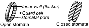
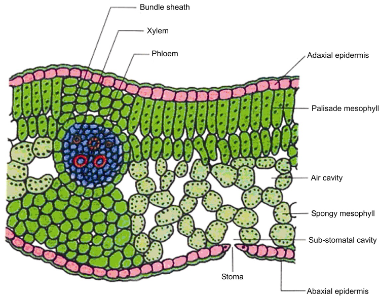
Carbondioxide: Terrestrial plants obtain carbon dioxide from the atmosphere through the small openings present on leaves called as stomata. `Stomata' are the small pores present on the surface of leaves they help in exchange of gases and water. Stomatal opening is guarded by the presence of guard cells (kidney shaped). Aquatic plants obtain CO2 dissolved in water through their general body surface so they perform more photosynthesis than terrestrial plants.
- Water: Plants absorb water from the soil by the process of osmosis. This water is transported to leaves by a special type of tissue called as xylem.
⇒ Plants utilize carbon dioxide during photosynthesis, the intensity of light at which amount of CO2 used during photosynthesis becomes equal to the amount of CO2 released during respiration by plants is called as Compensation point.
⇒ Compensation point occurs at low light intensity that is during morning and during evening hours.
Site of Photosynthesis:
Site of photosynthesis is different in prokaryotes and eukaryotes.
⇒ In prokaryotes: Photosynthesis occurs in lamellar chromatophores.
⇒ In eukaryotes: Photosynthesis occurs in chloroplast.
⇒ Exception: Fungi (It lacks chlorophyll so no photosynthesis occurs here)
⇒ In higher plants chloroplast is the main site of photosynthesis.
⇒ Chloroplast is also called as green plastid.
⇒ Plastid was first observed by Haeckel.
⇒ Plastids are of 3 different types on the basis of pigments present in them:-
(i) Leucoplast: White in colour, found in underground parts, lacks any coloured pigment.
Helps in storage of protein (Aleuroplast), oil (Elaioplast), starch (Amyloplast)
(ii) Chromoplast: Colour other than green, found in aerial parts of the plants.
(iii) Chloroplast: Contain green pigment, called as chlorophyll.
⇒ Chloroplast was discovered by Schimper.
⇒ Number of chloroplasts is variable in different species of plants.
⇒ In lower plants like algae they are 1 or 2 in number.
⇒ In higher plants their number varies from 40 -100 per palisade cell or more.
⇒ Chloroplasts also have variable shapes, for example cup shaped, ribbon shaped etc. in algae while it is discoidal in higher plants.
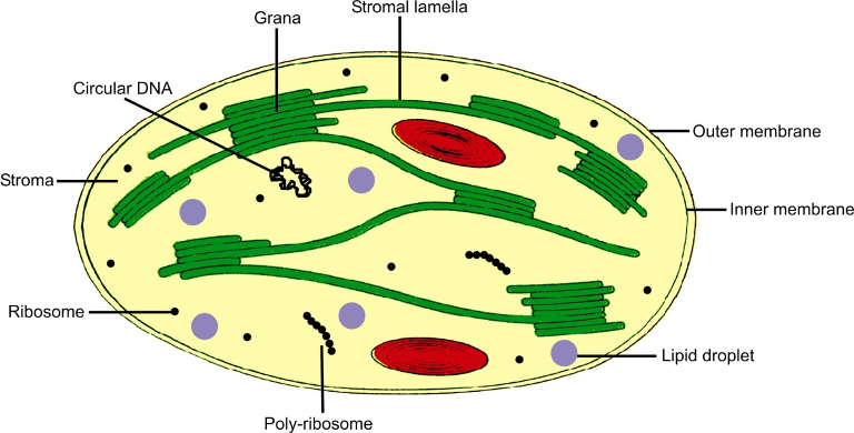
A typical structure of chloroplast is a double membranous structure having two parts:
(i) Grana: It is a lamellar system consisting of stacks of granum lamella each bounded by a membranous box called as thylakoid. They are 40 -60 per cell. Number of thylakoid per grana is 50 or more .Chlorophyll molecules are found inside the thylakoid membrane where they trap solar energy in the form of small energy packets called `photons 'or ' quanta'. Grana are interconnected to each other by a channel called as stroma lamellae or Fret's channel.
(ii) Stroma: It is a non pigmented proteinaceous matrix in which grana remain embedded It contains enzymes for dark reaction.
Mechanism of Photosynthesis:
(i) Light reaction:
› It is also called as photochemical process.
› It was discovered by 'Robert Hill' therefore it is also called as Hill’s reaction.
› Site: Grana of chloroplast.
› Raw materials: Light and water.
› Regulation: This process is regulated by chlorophyll molecules.
› It consists of 3 steps:
Photo excitation of chlorophyll molecule: During this process chlorophyll molecule receives sunlight in the form of small energy bundles called as photons and become excited to higher energy level.
Photolysis: It is also called as photo-oxidation of water; this takes place in presence of Mn+2 and CI- ions.
2H2O → 4H+ + O2 + 4e-
O2 is liberated as by product and H+ ions are used for reduction of NADP
2NADP +4H+ → 2NADPH2
Photophosphorylation: During this process ATP are produced. It takes place in quantasomes. Each quantasome contains about 230 to 300 chlorophyll.
MG+2 ions and inorganic phosphate is required to convert ADP→ ATP,
ADP + P (inorganic)→ ATP
(ii) Dark reaction:
- It is also called as thermo chemical reaction.
- It was discovered by Melvin Calvin and Benson therefore it is also called as
Calvin cycle
Site = stroma of chloroplast
- Raw materials: They require CO2, NADPH2, ATP and Enzymes.
- Regulated by: Light .reaction and enzymes.
- It involves three basic steps:
(i) Carboxylation: in this step CO2 is captured by CO2 acceptors like RUBP (C3 Plants) PEP (C4Plants) with the help of carboxylase enzyme i.e. RUBISCO & PEPCO respectively.
(ii) Synthesis: In this phase captured CO2 is assimilated into glucose in the presence of phosphatase and isomerase enzymes and RUBP is regenerated back.
(iii) Regeneration of RUBP
FACTORS AFFECTING PHOTOSYNTHESIS:
Light:
Normally plants utilize sunlight but marine algae can perform photosynthesis even in the moon light. Plants can also perform photosynthesis in the artificial lights.
- Highest rate of photosynthesis: Red light
- Minimum photosynthesis: Green light
- Very high light intensity can cause reduction in the rate of photosynthesis by causing
(i) Decrease in transpiration rate
(ii) Denaturation of chlorophyll molecule.
Temperature:
Optimum range = 25° C to 30° C
It ranges from 10 ° – 40° C
In some forms like algae of hot spring → 60° - 70° C is normal
Carbon dioxide:
It is the first limiting factor, 0.03 – 0.1 % is present in the atmosphere.
Concentration of CO2 → rate of photosynthesis. Above 0.9% → Rate
Between 0.1 to 0.9 %, it is constant and it is called as saturation point.
Oxygen:
O2 acts as competitive inhibitor of CO2. Over concentration of O2 stops photosynthesis.
Chlorophyll:
Chlorophyll content is directly proportional to rate of photosynthesis. No photosynthesis occurs in etiolated cells; in variegated leaves it occurs only at places where chlorophyll is present.
SIGNIFICANCE OF PHOTOSYNTHESIS:
Photosynthesis is a boon to the nature and to the human beings. It has following significance:
(i) Production of food material
(ii) Atmospheric control and purification of air
NUTRITION IN ANIMALS
⇒ Animals have highly evolved digestive mechanism that includes two basic components
⇒ Alimentary canal: Long, hollow, tubular structure consisting of various organs for digestion.
⇒ Digestive glands: They secrete enzymes / hormones which help in digestion.
⇒ Digestion in animals consists of following steps:
Ingestion : The process of intake of food.
Digestion : It is the breakdown of large and complex molecules into simpler, smaller and soluble forms.
Absorption : Taking up of the digested food through intestinal wall to blood.
Assimilation : In this process absorbed food is taken by body cells.
Egestion : The process by which undigested matter is expelled out.
⇒ Digestive system is regulated by various hormones secreted by some endocrine glands.
⇒ Alimentary canal was first of all developed in the phylum Platyhelminthes but only mouth was present in them
⇒ Coiled and well developed alimentary canal was developed in annelida till mammals.
NUTRITION IN LOWER ANIMALS:
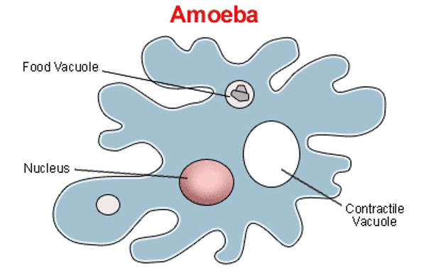
Nutrition in Amoeba:
Nutrition in amoeba is holozoic. Thus, solid food particles are ingested which are then acted upon by enzymes and digested. It is an omnivore, feeding on both plants and animals. Its diet includes bacteria, microscopic plants like the diatoms, minute algae, microscopic animals like other protozoa, nematodes and even dead organic matter.
Since it is a unicellular organism, amoeba does not have any specialised structure or organ for the process of nutrition. It takes place through the general body surface with the help of pseudopodia.
Mechanism of Nutrition Ingestion
The food is ingested at the point where it comes in touch with the cell surface with the help of pseudopodia. Pseudopodia engulf the food into the cytoplasm. The process of ingestion takes about two minutes.
Some methods of ingestion reported in amoeba are:
Circumvallation - When the prey is active, a food cup is formed with the help of pseudopodia.
Circumfluence:
When the prey is inactive and the amoeba rolls over it.
Import - The food passively sinks into the body on contact.
Invagination
Pseudopodia secretes a sticky and toxic fluid which adheres and kills the prey. It is then taken in by invagination.
Pinocytosis
Also called cell drinking. There are pinocytosis channels at certain points through which the cell ingests the food.
Digestion in Amoeba
Digestion in amoeba is intracellular taking place within the cell. The food taken in remains in a food vacuole or gastric vacuole formed by the cell membrane and small part of the cytoplasm. The vacuoles are transported deeper into the cells by cytoplasmic movements. Here they fuse with lysosomes that contain enzymes. Two enzymes amylase and proteinase have been reported. Thus, amoeba can digest sugars, cellulose and proteins. Fats, however, remain undigested.
The contents of the vacuole become lighter and the outline of the vacuole becomes indefinite indicating that the digestion is complete.
Absorption
Since the food on digestion is converted into liquid diffusible form, it is readily absorbed by the cytoplasm. The vacuole becomes progressively smaller as the food is absorbed by diffusion.
Assimilation
All the parts of the cell get the nutrients by the cyclic movement of the cytoplasm called the cyclosis. These nutrients are used to build new protoplasm. In this manner the food is assimilated.
Egestion
The egestion takes place by exocytosis. There is no particular point from which the egestion takes place. As the amoeba moves forward, the undigested matter is shifted to the back and eliminated as food pellets through a temporary opening formed at any nearest point on the plasmalemma.
Nutrition in Grasshopper:
⇒ It has a well developed digestive system having an alimentary canal and digestive glands.
⇒ The various organs of digestive system of grasshopper are
Mouth → Oesophagus → Crop → Gizzard → Stomach → Ileum → Colon → Rectum.
⇒ Glands associated with it are:
(i) Salivary glands
(ii) Hepatic caeca
⇒ Digestive system of a grasshopper can be divided into three parts.
(i) Foregut : mouth to gizzard
(ii) Midgut : gizzard to ileum (actual stomach)
(iii) Hindgut : stomach to anus.
⇒ The process involves:-
(A) Ingestion: It feeds on green leaves so it takes food through its mouth with the help of it's forelegs and mouth parts.
(B) Digestion:
(a) It starts from the mouth.
(b) A pair of salivary glands secretes saliva and release it into the mouth through the salivary duct.
(c) Saliva mixes with food and lubricates and softens the food.
(d) Digestion of starch begins here.
(e) This slightly digested food enters into the crop through a food pipe i.e. oesophagus.
(f) Crop stores the food temporarily.
(g) Now the food moves to gizzard. Here it is finally crushed and masticated and then moves to stomach.
(h) In stomach hepatic caeca releases its secretions in the form of digestive enzymes, thus the food is then completely digested at this site.
(C) Absorption: The digested food moves to small intestine (ileum) and absorbed through its walls.
(D) Assimilation: Nutrients are assimilated whenever required by the cells for the fulfillment of the growth, energy and repair of the body.
(E) Egestion: Undigested food is then passed through hindgut (where H2O is absorbed) and expelled out through anus in the form of elongated dry faecal pellets.
⇒ The excretory organ of the grasshopper is malphigian tubules present at the junction of hindgut and midgut.
Nutrition in Human Beings:
Human beings are heterotrophic omnivorous organisms. They obtain their food from plants, animals and their products by holozoic mode of nutrition.
Ingestion: Process of taking food inside the body through mouth.
Digestion: Process of conversion of complex food particles into simpler ones by the action of enzymes.
Absorption: Process of diffusion of soluble food particles into the blood.
Assimilation: Process of utilization of absorbed food by the body cells.
Egestion: Process of removal of undigested food from the body.
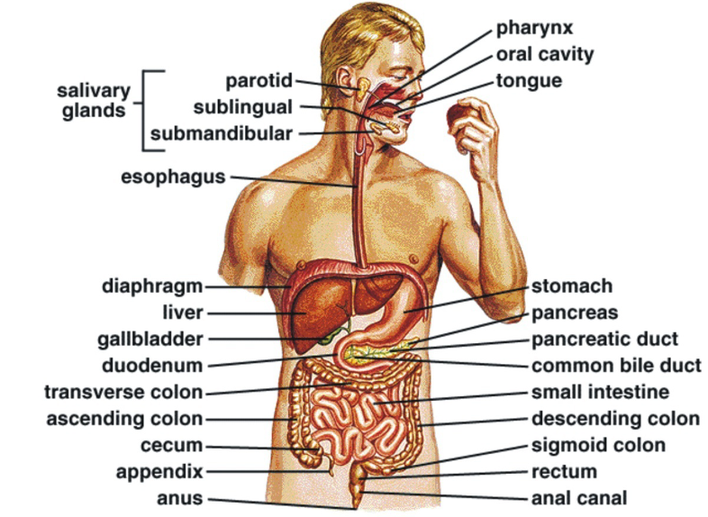
The organs, which take part in ingestion, digestion, absorption, assimilation and egestion together, form the digestive system.
The various steps of nutrition in human beings are:
Mouth:
It is a small slit through which food is ingested.
Buccal Cavity:
Mouth opens into a chamber called as buccal cavity. Roof of buccal cavity is called hard palate. At the floor of this cavity thick muscular structure is present called tongue. It helps in chewing, swallowing, tasting and speaking. Tongue has various types of papilla having taste buds.
⇒ Jaws present in buccal cavity are provided with four different types of teeth:
(i) Incisors : For cutting
(ii) Canines : For tearing
(iii) Premolars : For grinding
(iv) Molars : For grinding
⇒ Dental formula of humans:
(A) Milk teeth → These are temporary, arise at 6 - 11 month age, 20 in number
half upper jaw / half lower jaw = 2102/2102
(B) Permanent teeth → arise at 6 -12 years, 32 in number
half upper jaw / half lower jaw = 2123/2123
⇒ Three pairs of salivary glands are found in mouth which releases their secretions into the buccal cavity. They secrete salivary amylase for starch digestion. So digestion of starch starts from mouth.
Pharynx: The buccal cavity opens into a funnel shaped vertical canal. It is the common passage for food and air. It opens into oesophagus.
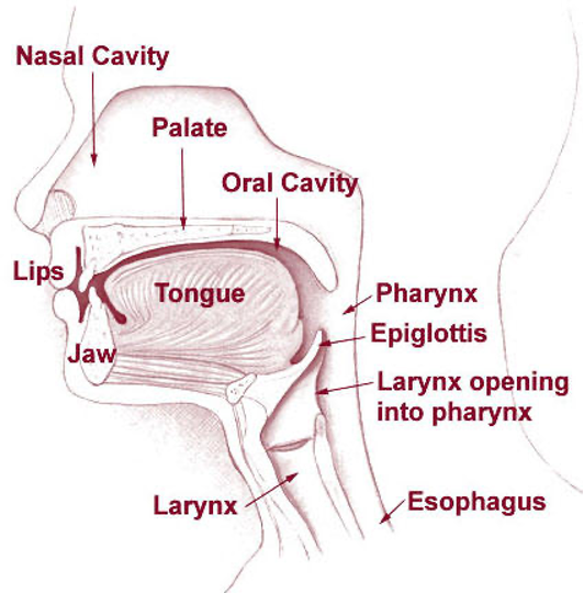
Oesophagus: Also called as food pipe. It leads the food from mouth to stomach. Oesophagus has highly muscular walls, no digestion occurs here.
Stomach: It is a large muscular J-shaped organ which expands when food enters it. The muscular walls of the stomach help in mincing the food thoroughly with more digestive juices. The stomach has branched and tubular glands present on its wall (gastric glands). The secretions of these glands are collectively called gastric juice.
The other functions of stomach include:
⇒ Storage of food.
⇒ Mechanical churning of food.
⇒ Regulation of the flow of food into the small intestine: From stomach, the food enters the small intestine. The exit of food from the stomach is regulated by a sphincter muscle which releases it in small amounts into the small intestine.
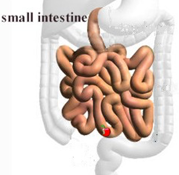
Small Intestine: From the stomach the food is moved to the small intestine. It is a very long tube (7 mt. long and 2.5 cm. diameter) which originates from the distal end of the stomach and extends to the large intestine. The small intestine is the longest part of the alimentary canal and is greatly coiled and twisted. The small intestine is subdivided into three sections: the duodenum, the jejunum, and the ileum.
The duodenum (about 10 inches ) part of the small intestine is the main seat of digestion in the gut. The acidic chyme in the duodenum receives the bile secreted from the liver, the pancreatic juice secreted from the pancreas and the intestinal juice from the glands of the intestinal wall. Bile is a yellowish-green in colour, bitter in taste, slightly alkaline fluid secreted from the liver. Bile being alkaline in nature neutralises the acidic chyme. Bile emulsify fat into microscopic droplets and thus helps in the digestion and absorption of fat. So bile is called digestive juice though it does not contain enzyme. Again the pancreas, a large gland located below the stomach, secretes pancreatic juice into the duodenum through the pancreatic duct. There are three enzymes namely amaylase, pancreatic lipase and trypsin, in pancreatic juice that break down carbohydrates, fats, and proteins respectively. Glands of intestine are present in the mucous layer of the intestinal wall. These glands secrete intestinal juice, which contains various enzymes. The enzymes present in it finally convert the carbohydrates into glucose, proteins to amino acids and fats into fatty acids and glycerol.
The jejunum is about 8.2 feet (2.5 meters) long. The digested carbohydrates, fats, proteins, and most of the vitamins, minerals, and iron are absorbed in this section. The inner lining of the small intestine is composed of up to five million tiny, fingerlike projections called villi. The villi increase the rate of absorption of nutrients into the bloodstream by greatly increasing the surface area of the small intestine.
The ileum, the last section of the small intestine, is the longest, measuring 11 feet (3.4 meters). Certain vitamins and other nutrients are absorbed here.
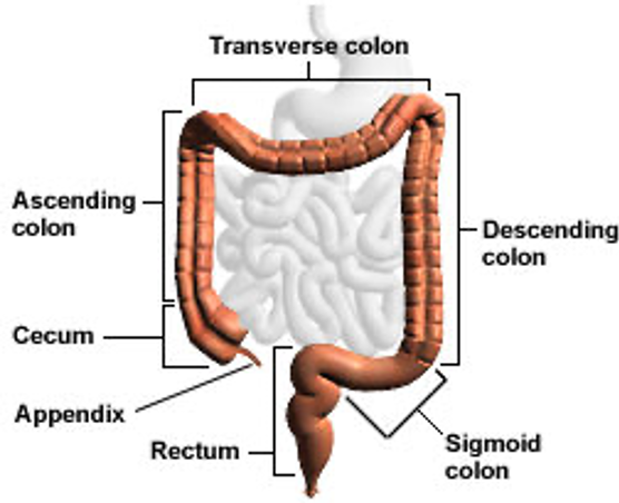
Large Intestine: Small intestine opens into large intestine from where the undigested food material is passed to anus through rectum. It is divided into three parts:
(i) Caecum (ii) Colon (iii) Rectum
Digestive Glands:
(i) Salivary glands: 3 pairs of salivary glands are found in mouth cavity. It helps in chemical digestion. They secret an enzyme called salivary amylase or ptyalin. It helps in digestion of starch.
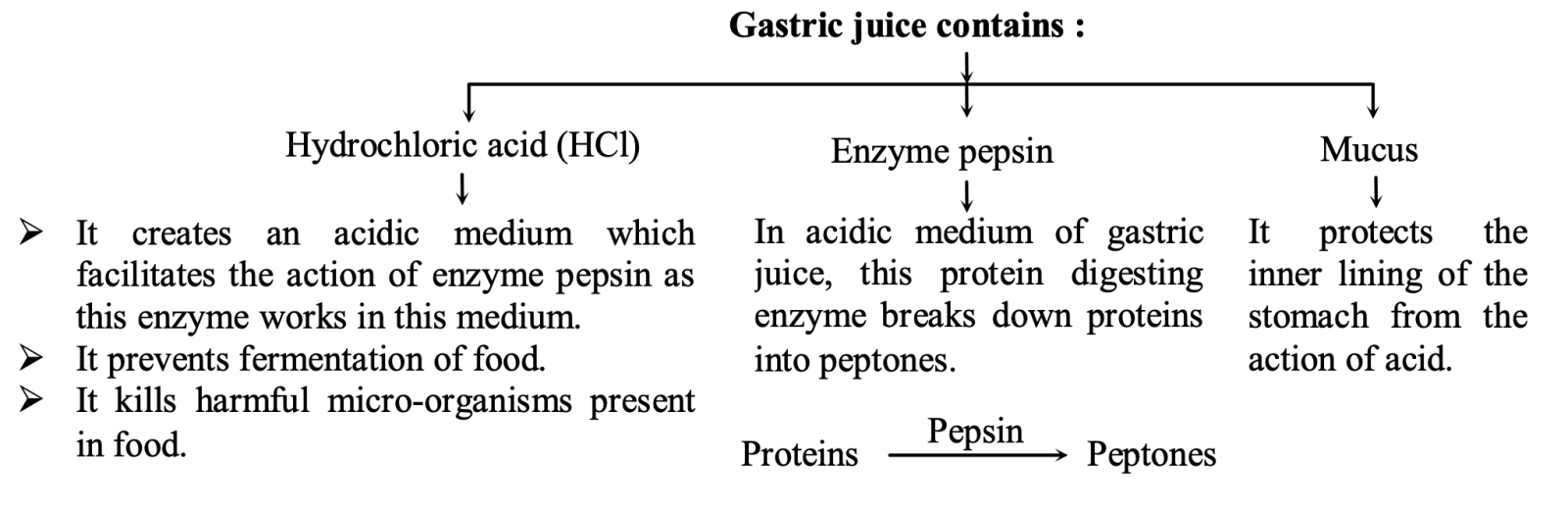
(ii) Gastric glands: Present in stomach. They secrete hydrochloric acid, protein digesting enzymes and mucus.
(iii) Liver: It is the largest gland, secretes bile into the small intestine. Bile contains bile juice and bile pigments. Bile is alkaline in nature and it is temporarily stored in gall bladder and helps in digestion of fats, it also helps in absorption of fats.
(iv) Pancreas: It lies parallel to and below the stomach. It secretes pancreatic juice into small intestine. Pancreatic juice contains trypsin and pancreatic amylase. Besides these 2 enzymes pancreas secretes 2 hormones also i.e. insulin and glucagon so it has both exocrine as well as endocrine functions. Both bile and pancreatic juice are released into the duodenum by a common duct.
INTESTINAL GLANDS: They secrete intestinal juice and mucus.
(a) Digestive System:
This system involves following processes:
(i) Ingestion: Intake of food is done through mouth, food is then chewed and masticated and sent to esophagus through pharynx by swallowing.
(ii) Digestion: Saliva secreted in buccal cavity starts digestion of starch into maltose. This partly digested food is then passed to stomach by oesophagus through peristaltic movement. Food is churned in stomach for about three hours and broken down into smaller pieces. Due to presence of hydrochloric acid, medium of stomach becomes acidic. In acidic medium protein digestive enzyme pepsin breaks down proteins into peptones. Gastric lipase is also secreted here which partially breaks down lipids.
- Secretion of gastric juice is stimulated by the sight, smell or thought of food.
- Now the partly digested food moves to small intestine i.e. in the duodenum. Duodenum receives the secretion from liver and pancreas through a common duct they are bile and pancreatic juice, and alkaline in nature. So the digestion and emulsification of fats occurs at this place. Bile juice also acts as a medium to provide alkalinity for action of pancreatic enzymes.
- Here in the duodenum fats are emulsified into fatty acids and glycerol by bile, remaining proteins are digested by trypsin and starch by pancreatic amylase.
-
This partially digested food now enters in the ileum where intestinal juice i.e."Succus entericus" is secreted. At this place digestion is completed.
Carbohydrates → Glucose
Proteins → Amino acids
Fats → Fatty acids and glycerol
(iii) Absorption: After digestion molecules are broken down into simpler water soluble forms now they are to be utilized, so they pass through the wall of small intestine which contains blood capillaries and enters into the blood. For absorption of fat lymph capillaries are present called as lacteals.
(iv) Assimilation: The process of utilization of food is called assimilation. The nutrients dissolved in blood are carried to all parts of the body where they are utilized.
(A) For building up and replacement of cells.
(B) For obtaining energy. This energy is released by the process of oxidation during respiration.
(v) Egestion: The undigested food is then collected in large intestine where water is absorbed and remaining waste is expelled out or egested through anus.
Enzymes: The enzymes are proteinaceous substance which plays an important role in digestion. The study of enzymes is enzymology and the term enzyme was coined by Kuhne in 1878.
General characteristics of enzymes:
- Enzymes are proteins.
- Enzymes are capable of promoting chemical reactions without losing their identity and therefore are termed biological catalysis.
- When the temperature increases the enzymatic activity increases till on optimum temperature is reached. When the temperature increases beyond the physiological range, enzymes get denatured and lose their activity.
- Even a minimum quality of an enzyme can bring about a chemical reaction.
- Normally, enzymes are destroyed above the optimum temperature. But certain enzymes like pepsin continue their proteolytic activity even at very high temperature.
- Enzymes are highly specific and they act only on a specific substrate.
7. The enzymatic activity may be accelerated by the presence of certain ions like calcium and magnesium.
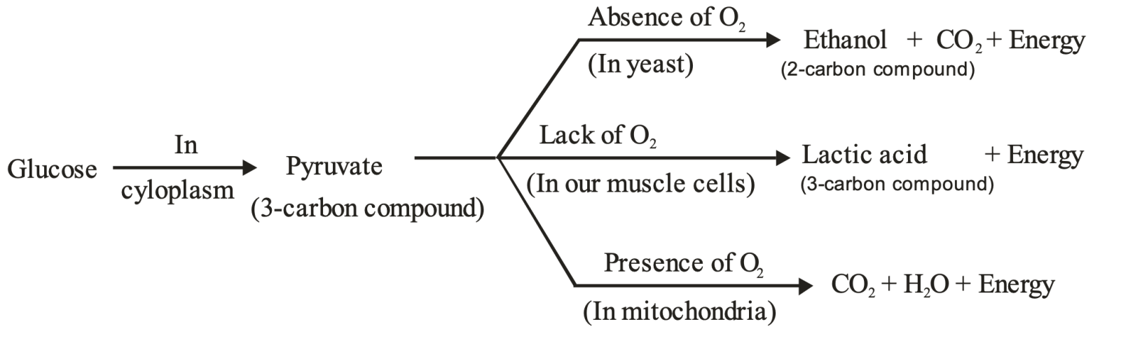
RESPIRATION
The sum total of all the vital activities is called as metabolism. Vital activities refer to all the physiochemical activities of a cell. It has two aspects:
(i) Anabolism: It includes metabolic processes by which complex cellular compounds are synthesized from simpler compounds, e.g. Photosynthesis
(ii) Catabolism: It includes metabolic processes by which larger molecules are broken down into simpler molecules e.g. Respiration. Respiration is an important catabolic process responsible for the production of energy.
(a) Definition: The process by which assimilated food is oxidised and energy is released is called as respiration. In this process oxygen from air is taken in, this oxygen reacts with food molecules present in the body cells and burn them slowly to release energy. This energy is stored in the form of ATP molecules inside the cell for further use and the waste products i.e. CO2 and H2O are eliminated out of the body. It is called as aerobic respiration.
TYPES OF RESPIRATION:
Aerobic Respiration: Some organisms use oxygen to break-down glucose (food) in cells completely into carbon dioxide and water. Aerobic organisms need to ensure that there is sufficient intake of oxygen.

Anaerobic Respiration: Organisms like yeast do not use oxygen and break-down of glucose is not complete resulting in break-down of glucose into ethanol, carbon-dioxide and release of energy.
Similarly, during vigorous exercise in our body muscles, anaerobic respiration takes place resulting in formation of lactic acid and energy.

Difference between aerobic and anaerobic respiration:
|
S.No. |
Aerobic Respiration |
Anaerobic Respiration |
|
1. |
In this type of respiration Oxygen is needed. |
In this type of respiration Oxygen is not needed. |
|
2. |
In this type or respiration there is complete oxidation of nutrients and no production of any organic by-product. |
In this type or respiration there is incomplete oxidation of nutrients and no production of any organic by-product. |
|
3. |
In this type of respiration a large amount (686 Kcal.) or energy is released. |
In this type of respiration a large amount (54 Kcal.) or energy is released. |
|
4. |
The final product of Aerobic Respiration are Carbon Dioxide and Water. |
The final products of Anaerobic Respiration are Ethanol in medium of Anaerobic Respiration are Ethanol in medium of plant origin and Lactic acid in medium of animal origin. |
|
5. |
It occurs in two phases: (i) Krebs Cycle and (ii) Oxidative Phosphorylation |
It never occurs in two phases. |
|
6. |
It is a complex process. |
It is a simple process. |
|
7. |
It is occurs in the cytoplasm and the mitochondria. |
It entirely occurs in the cytoplasm. |
|
8. |
It is seen in most of the organisms which can obtain oxygen from water or atmosphere. |
It is seen in certain bacteria. Yeast and other fungi, end oparasites and animal muscle cells. |
Steps of respiration
⇒ In all cases, the first step is the breakdown of glucose, a six-carbon molecule, into a three carbon compound called Pyruvate.
⇒ This break-down takes place in cytoplasm.
⇒ The break-down of pyruvate in presence of oxygen takes place in mitochondria resulting in release of energy. Hence, mitochondria are also known as power-house of the cell.
ATP : The energy currency of the cell:
⇒ It is a nitrogenous organic compound. The energy released during cellular respiration is used to synthesise, a molecule called ATP (Adenosine triphosphate) which is the energy currency of living organisms.
ADP+ (P) ADP ~ (P) = ATP P = phosphate
⇒ It is commonly called energy currency of the cell.
⇒ When the terminalphosphate linkage in ATP is broken using water, the energy equivalent to 30.5 KJ / mol is released.
⇒ ATP is used in body for muscle contraction, protein synthesis conduction of nerve impulses and all other activities.
Respiration:
Respiration in divided in three parts:
(i) Cellular respiration
(ii) Respiration in plants
(iii) Respiration in animals
Respiration in Plants:
- In plants exchange of gases takes place from leaves, stems and roots individually.
- Transfer of respiratory gases from one part to another is very less.
- Exchange of gases in plants occurs by simple diffusion.
(i) Respiration in roots:
- In young roots, the epidermal cells are extended to form root hair. These root hairs remain in direct in contact with the air present in between the soil particles. The oxygen from this air enters into the root hairs by simple diffusion and reaches to other cells of root for respiration.
- In older roots a protective layer of dead cells is present which have tiny openings called as lenticels. Diffusion of oxygen takes place through these pores and carbon dioxide is released out through the same.
(ii) Respiration in stem:
- In herbaceous plants, stem have small openings in their epidermal cells called as stomata, the oxygen from air enters through stomata and carbon dioxide is released from the same.
- In hard and woody stems of big plants and trees, lenticels are present in place of stomata through which exchange of gases takes place.
(iii) Respiration in leaves:
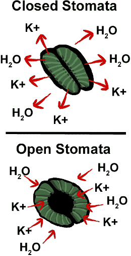
- Surface of leaves possess numerous tiny pores called as stomata in their epidermal cells; exchange of gases takes place through stomata. Massive amounts of gaseous exchange takes place in the leaves through stomata for the purpose of photosynthesis. But it is important to note here that exchange of gases occurs across the surface of stems, roots and leaves as well. Since large amounts of water can also be lost through these stomata, the plant closes these pores when it does not need carbon dioxide for photosynthesis. The opening and closing of the pore is a function of the guard cells. The guard cells swell when water flows into them, causing the stomatal pore to open. Similarly the pore closes if the guard cells shrink.
- An experiment to show that Plants take oxygen and evolve carbon dioxide during respiration:
Experiment: To demonstrate that the plants take oxygen and evolve carbon dioxide during respiration set the apparatus according to figure by taking KOH in U-tube, lime-water in two wide mouth bottles, one potted plant, bell jar and black-cloth. During day time the potted plant is covered with black-cloth to check photosynthesis. Make the apparatus airtight and start the aspirator. After some time, the lime water of second bottle turns milky. The explanation for this is that when the water comes out from aspirator, the atmosphere air enters into the apparatus through the second end and passes through the U-tube containing caustic potash into the tube containing lime water. The caustic potash absorbs the CO2 of the air. Thus, CO2 free air reaches into the lime water so it does not turn milky. It indicates the air does not contain even trace of CO2. When this air reaches into the lime water of second tube through a bell jar having potted plant covered with black cloth to check photosynthesis, it turns milky. It proves that CO2 is evolved during respiration.
RESPIRATION IN ANIMALS
Animals have evolved different organs for the uptake of O2 and release of CO2. However in unicellular organisms no specific organ is there for exchange of gases because the entire surface of the organism is in contact with the environment.
- Aquatic animals use the O2 dissolved in water. Since the amount of O2 in water is fairly low as compared to the amount of O2 in the air, the rate of breathing in aquatic organisms is much faster than that seen in terrestrial organisms.
- Fishes, take in water through their mouths and force it past the gills where the dissolved O2 is taken up by blood.
- Terrestial organisms use the O2 in the atmosphere for respiration. This O2 is absorbed by different organs in different animals.
Type of respiration Organs involved Example
- Cell surface respiration General body surface Amoeba. Paramecium
- Tracheal respiration Trachea and tracheoles Insects
- Branchial respiration Gills Fishes
- Cutaneous respiration Skin Frog
- Pulmonary respiration Lungs Amphibians, reptiles, birds
- Buccal respiration Buccal cavity Frog
Some important characteristics of respiratory organs of animals are:
- They have large surface area to get enough oxygen.
- They have thin walls for easy diffusion and exchange of gases.
- They have rich blood supply for transport of respiratory gases.
Respiration in Amoeba:
The taking in of oxygen and removal of carbon dioxide – the gases simply diffuse through the whole of amoeba's permeable surface. Soluble nitrogen-containing wastes, produced by the amoeba's chemical activities are excreted in the same way.
Respiration in Earthworm: In organisms like earthworm and leech exchange of gases occurs through their skin as their skin is very thin and moist. It is rich in blood supply so the oxygen is absorbed by moist skin of earthworm and is transported to all the cells of body through blood. The carbon dioxide from body cells diffuses into the blood and expelled out through skin.
Respiration in Fish:
- In fish exchange of gases occurs through gills so the respiration is said to be branchial.
- Gills are present on both the sides of its head; they are covered by gill covers.
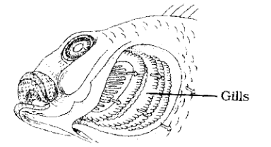
- During breathing fish takes in water through its mouth and pass it over the gills, the oxygen present in water extracted by gills and water is removed out through gill slits. This oxygen is now absorbed by blood and carried to all parts of the body and at the same time carbon dioxide is released into the blood and comes back to the gills and is expelled out into the surrounding water.
- Same type of respiratory pattern is followed in some other aquatic organisms like prawns.
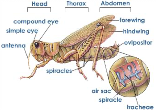
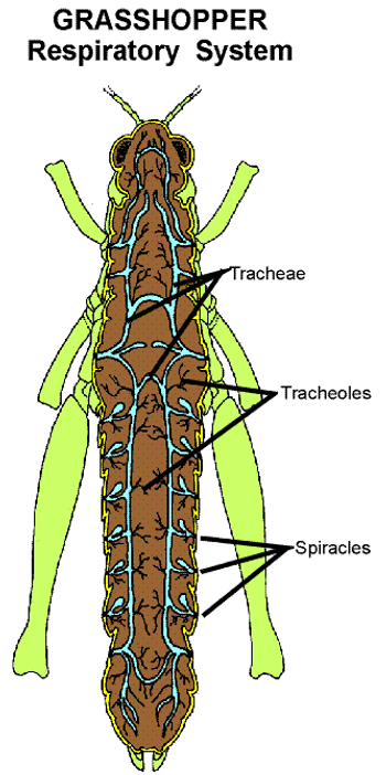
Respiration in Grasshopper:
The respiratory system in insects is called the tracheal system. It involves the diffusion of oxygen directly from the atmosphere into the air-filled tubes. Thus, the diffusion is through air and hence, is more efficient than the diffusion through water (300,000 times more) or tissues (1,000,000 times more).
In grasshopper, the tracheal system consists of 10 pairs of spiracles, located laterally on the body surface. Of these, 2 pairs are thoracic and 8 pairs are abdominal. The spiracles are guarded by fine hairs to keep the foreign particles out and by valves that function to open or close the spiracles as required. The spiracles open into small spaces called the atria that continue as air tubes called the tracheae. The tracheae are fine tubes that have a wall of single layered epithelial cells. The cells secrete spiral cuticular thickenings around the tube that gives support to the tubes.
Comparison between respiration in animals and plants:
|
S.No. |
Respiration in Animal |
Respiration in Plants |
|
1. |
Animal performs respiration as a single unit. |
All parts of plant (like root, stem, leaves) perform respiration individually. |
|
2. |
Respiratory gases are usually transported to long distances |
There is little transport of respiratory gases from one part of the plant to other. |
|
3. |
Respiration occurs at faster rate. |
It occurs at slower rate. |
Respiration in Humans:
Human respiratory tract
(i) External nostrils: First part of respiratory system. It opens into nasal cavity and is meant for inhalation of air from outside.
(ii) Nasal cavity: This cavity is separated from oral cavity by means of a hard and bony palate. It is lined by ciliated columnar epithelial cells that are rich in mucus, it brings about warming, moistening and sterilization of air It contains hair and mucus which entrap the dust particles.
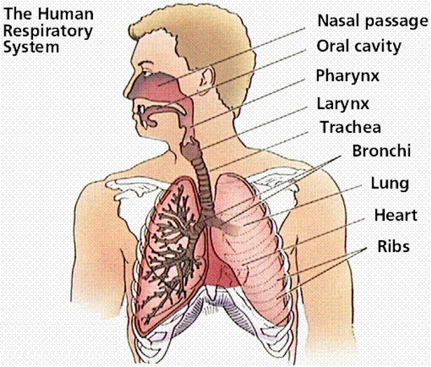
(iii) Internal nares: Nasal cavity opens into it and it leads to pharynx.
(iv) Pharynx: It is a common part between both alimentary canal and respiratory system.
(v) Larynx: It is an enlarged part of trachea which is also called as `voice box'. It produces voice by passage of air between vocal cords. It contains four different types of cartilages among them a 'c' shaped thyroid cartilage protruding out in neck region is called Adam's apple.
(vi) Trachea: also called wind pipe. It is 10-12 cm long tube. Its walls are supported by 16 - 20 'c' shaped cartilaginous rings which prevent them to collapse when air is absent in them.
(vii) Bronchi: Trachea is branched into two bronchi left and right each of which enters into the lungs.
(viii) Lungs: These are two light weight spongy pouches covered by a membrane called Pleura. Bronchi are further branched into several bronchioles, at the end of bronchioles alveolar sacs or alveoli are present which are rich in blood capillaries and thin walled.
(ix) Diaphragm: It is a sheet of muscles that lies below the lungs and separates thoracic cavity from abdominal cavity.
Mechanism of Breathing:
Mechanism of breathing involves two phases.
Inspiration (Inhalation): It is the process by which fresh atmospheric air enter into the lungs (alveoli) via respiratory tract. The diaphragm contracts and becomes flat expanding the chest cavity. The intercostal muscles contract, ribs coming outwards. This further enlarges the chest cavity, lowering the pressure inside lungs. Therefore air rushes in.
Expiration (exhalation): It is the process by which foul air is expelled out of the lungs. The diaphragm relaxes and coming in its normal arched position, compressing the lungs. Intercostal muscles relax and move inwards. This reduces the volume of chest cavity which is already full of air. This forces the air out.
Inhalation → Lifting of Ribs + Flat Diaphragm → Increase in Volume of Chest Cavity → Air is sucked inside the Nostrils → Alveoli and Vice Versa for Breathing out.
During breathing cycle, when the air is taken in and let out, the lungs always contain a residual volume of air so that there is sufficient time for O2 to be absorbed and for the CO2 to be released.
The blood brings carbon dioxide from the rest of the body for release into the alveoli, and the oxygen in the alveolar air is taken up by blood in the alveolar blood vessels to be transported to all the cells in the body.
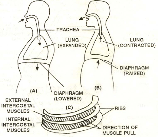
Mechanism of breathing
Exchange of Gases CO2 and O2 between blood and tissues:
When the body size of animals is large, the diffusion pressure alone cannot take care of oxygen delivery to all parts of the body. Instead respiratory pigments take up oxygen from the air in the lungs and carry it to tissues which are deficient in oxygen before releasing it.
In humans, respiratory pigment haemoglobin is present in the RBC which has very high affinity for O2. Solubility of CO2 is more as compared to O2 in water and hence is mostly transported in dissolved form in our blood. Carbon mono-oxide when binds with haemoglobin a stable compound carboxy-haemoglobin is formed which can cause death as no haemoglobin is left for transport of O2.
Air in Alveoli Blood Vessels Blood RBC Respiratory Pigment (Haemoglobin) Oxygen Links with Hb (high pressure of O2) O2 is released in tissues from Hb (Low O2 Pressure in Tissues) High CO2 in Tissues CO2 Released into Blood Blood Vessels in Alveoli CO2 Released out Through Nostrils.
TRANSPORTATION
All living bodies need nutrients and oxygen in every cell of its various tissues to sustain life. The transport of different materials and gases is essential both in plants and animals. Unicellular organisms e.g. Amoeba and Paramecium do not require the transport of any material. These are in direct contact with their surroundings from where they obtain these nutrients. These substances are distributed in the cytoplasm due to the streaming movements of cytoplasm called as cyclosis. They exchange gases from the external environment directly by diffusion due to the difference in the concentration in and outside their body. In higher organisms both plants and animals, digested food, oxygen, hormones, waste nitrogenous substances etc. are to be carried from one place to the other. So transportation of materials is essential. It is done through circulatory system.
TRANSPORTATION IN PLANTS:
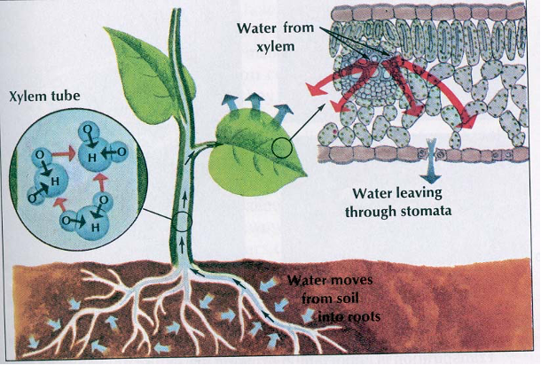
For plants, the soil is the nearest and richest source of raw materials like nitrogen phosphorus and other minerals which are required for photosynthesis as well as other metabolic activities. The absorption of these substances therefore occurs through the part in contact with the soil, namely roots. If the distances between soil-contacting organs and chlorophyll containing organs are small, energy and raw materials can easily diffuse to all parts of the plant body. Diffusion processes will not be sufficient to provide raw material in leaves and energy in roots. A proper system of transportation is therefore essential in such situations.
Energy needs differ between different body designs. Plants do not move, and plant bodies have a large proportion of dead cells in many tissues. As a result, plants have low energy needs, and can use relatively slow transport systems.
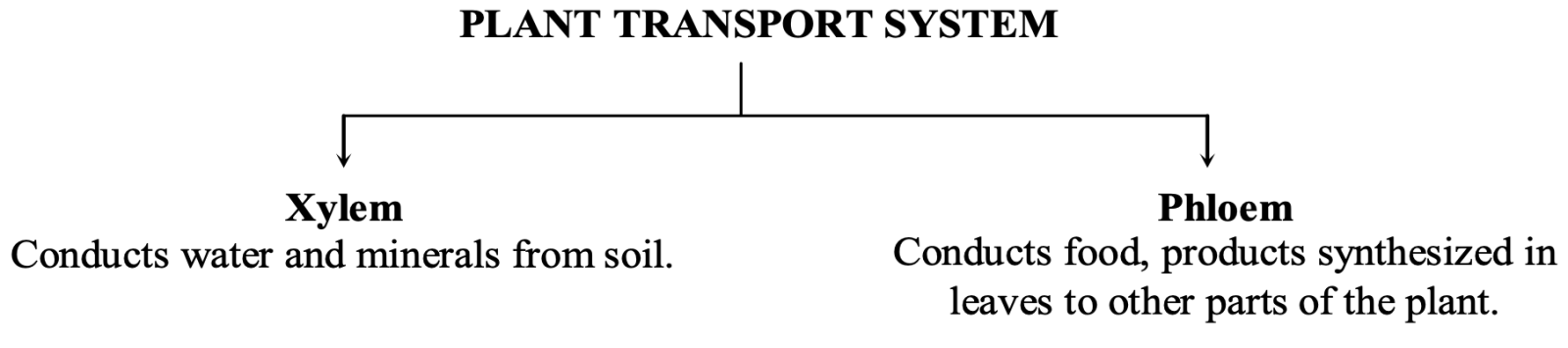
Plant transport systems will move energy stores from leaves and raw materials from roots. These two pathways are constructed as independently organized conducting tubes.
Translocation:
Phloem translocates the manufactured food sugar or starch from the leaves to the different parts of the plant including the roots.
Transpiration:
Most of the water absorbed is lost through the aerial parts of the plant into air by a process called "transpiration". Two percent of total water absorbed is used up in various metabolic activities in the plant body. Transpiration is the loss of water from the living tissues of the aerial parts of the plant in the form of water vapours. There are three types of transpiration:
- Cuticular transpiration (through cuticle)
- Lenticular transpiration (through lenticels)
- Stomatal transpiration (through stomata)
Importance of transpiration:
(A) It controls the rate of absorption of water from the soil.
(B) It is responsible for ascent of sap.
(C) It regulates the temperature of the plant.
(D) Mostly water absorbed by roots is lost by transpiration without serving any purpose. The energy spent by the plants in transpiration is wasted. So transpiration is a necessary evil.
Difference between transport by xylem and phloem:
|
Properties |
Phloem |
Xylem |
|
Function: |
Transportation of food and nutrients from leaves to storage organs and growing parts of plant. |
Water and mineral transport from roots to aerial parts of the plant. |
|
Movement: |
Bidirectional (Moves up or down the plant's stem from "source to sink") |
Unidirectional (Moves up the plant's stem) |
|
Occurrence: |
Roots, stems and leaves |
Roots, stems and leaves |
|
Additional Functions: |
Forms vascular bundles with xylem |
Forms vascular bundles with phloem and gives mechanical strength to plant due to presence of lignified cells. |
|
Structure: |
Tubular with soft walled cells |
Tubular with hard walled cells |
|
Elements: |
Sieve tubes, companion cells, phloem parenchyma, bast fibers, intermediary cells |
Tracheids, vessel elements, xylem parenchyma, xylem sclerenchyma |
|
Nature of tissue: |
Living tissue |
Non living tissue at maturity |
TRANSPIRATION COHENSION THEORY:
The main loss of water is through stomatal transpiration. Turgor pressure in the mesophyll cells of the leaf forces water outwards through the cell wall. Water evaporates from the surface of the cells into the air spaces of the spongy tissues and then passes into the outer atmosphere through the pores or stomata. The cell sap of mesophyll cells becomes concentrated by losing water and causes a drop in turgor pressure. As a result water is sucked from adjoining mesophyll cells and ultimately from vascular tissues. This tension is transmitted all the way down to the unbroken column of water through the stem to the absorbing parts of the root. The molecules of the water show cohesion (mutual attraction) and molecules of water and vessel wall show adhesion (affinity for water). Due to these adhesive and cohesive forces, water column does not break but pulled upward by the force called as "transpiration pull". The whole process can be compared with a person (transpiration pull) pulling a bucket full of water (forces on water column) from a well with a rope (column of water due to cohesion).
TRANSPORTATION IN HUMAN
In human’s transportation of O2, nutrients, hormones, and other substances to the tissues, CO2 to the lungs and waste products to the kidney is carried out by a well-defined circulatory system.
The circulatory system comprises of :
- Blood vascular system
- Lymphatic system
Blood Vascular System:
The higher multi-cellular animals with higher metabolic rates possess a well developed blood vascular system. This system helps in the quicker supply of nutrients and oxygen to the body tissues and also in the rapid disposal of toxic waste materials and carbon dioxide. The blood acts as the circulatory fluid. Blood vascular system consists of blood, blood vessels and heart.
- Blood: Blood is a specialized bodily fluid in animals that delivers necessary substances such as nutrients and oxygen to the cells and transports metabolic waste products away from those same cells. Blood is circulated around the body through blood vessels by the pumping action of theheart. In animals with lungs, arterial blood carries oxygen from inhaled air to the tissues of the body, and venous blood carries carbon dioxide, a waste product of metabolism produced by cells, from the tissues to the lungs to be exhaled.
- Plasma: The plasma consists of water (90% & above) inorganic and organic substances. In the plasma, RBCs, WBCs and blood platelets float. Inorganic salts (0.9%) are also present. The organic substances are glucose, amino acids, proteins, and hormones, digested and waste excretory products. The blood proteins (7%) are fibrinogen, albumin, globulin and prothrombin.
RED BLOOD CORPUSCLES (RBCS) OR ERYTHROCYTES:
These are one of the smallest cells in your body, they are round with a dent in the middle, we call this shape a Biconcave disc.The function of the red blood cells is to transport oxygen from the lungs to the body cells. A red protein called Haemoglobin, when the blood reaches the lungs, oxygen diffuses from the alveoli to the red blood cells and combines with haemoglobin forming an unstable compound called oxyhaemoglobin. When the blood reaches the body cells, the oxyhaemoglobin is easily split into oxygen and haemoglobin again, the oxygen diffuses through the blood plasma to the cells.
Red blood cells are fully adapted to their function by the following characteristics:
(i) Biconcave disc shape gives it large surface area to carry more oxygen.
(ii) Haemoglobin to combine with oxygen
(iii) No nucleus that takes up space.
WHITE BLOOD CELLS:
White blood cells are one of the substances floating in the blood plasma. They are completely different in function than red blood cells. White blood cells are part of the Immune System, they play a big role in protecting the body by killing bacteria which cause disease, also known as pathogens. White blood cells can be distinguished from red blood cells easily because they are much bigger, with a nucleus, and present in fewer amounts.
TYPES OF WHITE BLOOD CELLS:
Phagocytes:
They kill bacteria by engulfing them, taking them in the cell then kill them by digesting them using enzymes, this process is called phagocytosis.
Most white blood cells are the phagocyte type.
Lymphocytes:
Unlike phagocytes, lymphocytes have a large nucleus. They are produced in the lymph nodes (in the lymphatic system). Lymphocytes kill bacteria by secreting antibodies and antitoxins which kill the pathogens directly or make them easier to kill. Each pathogen could be killed by a certain type of antibody.
The Platelets:
Platelets are tiny cell fragments that prevent bleeding when the skin is cut, and stops bacteria from entering our systems through the wound. This works by blood clotting, when the skin is cut, some reactions take place that results in platelets producing a protein, this protein will change the fibrinogen (another soluble protein in the plasma) to insoluble fibrin. The fibrin forms long fibres that clot together blocking the cut, thus preventing any bleeding, this is called blood clotting.
Blood Plasma:
This makes up most of the blood. It is mostly water with some substances dissolved in it, these include carbon dioxide, hormones, food nutrients, urea and other waste products. The blood plasma transports substances from one place to another.
Functions of the blood:
- Transportation of R.B.C’s, W.B.C’s, oxygen, food nutrients, hormones, and waste products.
- Defence against disease, by white blood cells phagocytosis and production of antibodies.
- Supplying cells with glucose to respire and keep a constant temperature.
Blood Vessels (Vascular System):
This is a number tubes carrying blood away from and to the heart and other organs. The main types are Arteries, Veins and Capillaries.
Arteries:
Their function is to transport blood away from the heart to the lungs or other body organs.
The blood in the arteries always has a high pressure. The heart pumps the blood quickly into the arteries, resulting in the pressure, each time the ventricle of the heart contracts, the pressure in arteries increase, when the ventricle relaxes, the pressure falls. The lumen of arteries is also very narrow, adding to the pressure.
The structure is simple, beside the narrow lumen, the arteries have a strong thick wall to withstand the pressure. Their walls are also elastic and stretchable.
Brief description of characteristics of arteries:
- Transporting blood away from the heart
- Always in a high pressure
- Strong but stretchable walls
- Narrow lumen.
Veins:
Their function is to transport blood to the heart from the body.
The veins always always have a low blood pressure because by the time the blood with high pressure reaches the veins, it loses most of the pressure. This means that blood flows very slowly in veins, to help this, veins lie between muscles so that the blood is squeezed when the muscles contract.
They have a simple structure. Because they have a low pressure, they don’t need strong, thick walls like the artery, instead they have thin less elastic walls. Their lumen is much wider too. Veins have a unique feature that is valves. Because blood in veins flows slowly with a low pressure, there is a risk of a backflow, especially in veins that move blood upwards against gravity, like the ones in the leg. The valves ensure that the blood is always flowing in the direction of the heart. When the muscles squeeze the blood, the valves are open the let blood through, when muscles relax, valves close to prevent a backflow.
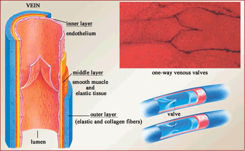
Brief description of characteristics of veins:
- They carry blood to the heart
- Always in a low pressure
- Thin less elastic walls
- Wide lumen Valves present.
| Artries | Veins | ||
| 1. | Blood flows away from the heart | 1. | Blood flows towards the heart |
| 2. | Posses thick elastic walls | 2. | Posses thin, not very elastic walls |
| 3. | Carry oxygenated blood except pulmonary artries | 3. | Carry deoxygenated blood except pulmonary vein |
| 4. | Do not posses valves except in the aorta | 4. | Have valves to prevent back flow of blood |
| 5. | Arteries are deeper in the flesh than veins | 5. | Veins are nearer the surface of the skin than arteries |
| 6. | Pulse is detectable | 6. | Pulse is usually not detectable |
| 7. | Have narrow lumen | 7. | Have wonder lumen |
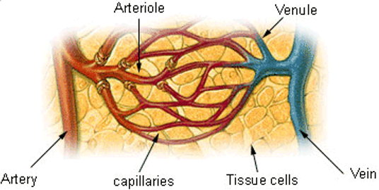
Blood Capillaries:
Blood capillaries are the smallest blood vessels in our systems. Their function is to get blood from the arteries as close as possible to the tissues in order to exchange materials with the cells, and to link arteries with veins.
When arteries come near and organ or a tissue, it divides into arterioles, these arterioles divide more into several blood capillaries that go through the tissue, this is when the exchange of oxygen and food nutrients with carbon dioxide and waste products such as urea take place by diffusion.
Blood capillaries are very well adapted to their jobs. They are one cell thick to reduce the diffusion distance of materials for faster diffusion. They also have pores in their walls between the cells, to allow the plasma to get out of the blood and become tissue fluid.
The Heart:
The heart is a pumping organ that is responsible for the movement of blood around the body. The function of the heart is to give the blood a push, keeping it flowing around the body all the time. That is why the heart is constantly working, if it stops for a minute, the other organs will not receive any oxygen or nutrients, thus the body fails and the person dies. The heart is located in the chest, the thoriac cavity between both lungs.
Structure:
The heart is hollow, it has 4 chambers. Two of them are atria and two are ventricles. One of each of these on each side. When looking at the diagram of a heart, notice that your right is the left side of the heart, and your left is the heart’s right, as if you are looking at your own heart on a mirror.
The sides of the heart are separated by a wall called septum. Each side contains an atrium (at the top) and a ventricle (at the bottom), there is a valve between the atrium and the ventricle in each side, it is called bicuspid valve in the left side and tricuspid valve in the right side. There are several blood vessels associated with the heart, these are:
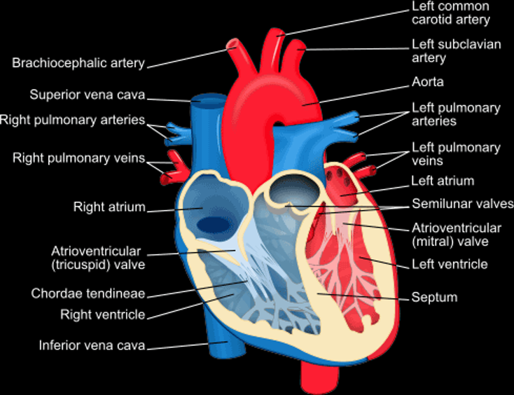
- The Pulmonary vein, it transports oxygenated blood from the lungs to the right atrium.
- The Aorta, which is the biggest artery in the body, it transports oxygenated blood from the heart to the rest of the body.
- The Vena Cava, the biggest vein in the body, it transports deoxygenated blood from the whole body to the heart.
- The pulmonary artery, it transports deoxygenated blood from the heart to the lungs.
Note that blood vessels entering the heart are veins, and the ones leaving the heart are arteries. The left side of the heart always contains oxygenated blood because it receives blood fresh from the lungs and pumps it to the body, the right side always contains deoxygenated blood because it receives is from the body. You can memorize this by the word LORD: Left Oxygenated – Right Deoxygenated.
The heart receives blood from the lungs at the left atrium and pumps it to the body from the left ventricle, then it receives it again from the body at the right atrium and pumps it to the lungs from the right ventricle. The red shows oxygenated blood and the blue shows deoxygenated blood.
The heart receives blood from the lungs at the left atrium and pumps it to the body from the left ventricle, then it receives it again from the body at the right atrium and pumps it to the lungs from the right ventricle. The red shows oxygenated blood and the blue shows deoxygenated blood.
Notice that the walls around the left ventricle are much thicker than the ones in the right ventricle. The reason for this is that because the left ventricle pumps blood to the whole body, so blood will travel a long distance, so it needs lots of muscles to contract and pump the blood more strongly.
However, the right ventricle pumps blood the lungs which are very close to heart, the blood does not need to be pumped very strongly.
Mechanism of the heart:
When the heart is being filled with blood (whether from the body or the lungs), this is called the diastole. When the heart is pumping the blood out of it (whether to the body or to the lungs), it is called the systole.
During diastole, the heart is getting filled with blood, the blood enters the atria first, the atria contract to force blood into the ventricles, both tricuspid and bicuspid valves are open to allow blood into the ventricles and the semilunar valves are shut. Once the ventricles get filled with blood, it is systole, the bicuspid and tricuspid valves get shut and semilunar valves are open, the ventricles contract strongly forcing the blood into the Aorta or pulmonary artery.
During diastole the semilunar valves are shut to keep the blood out of the arteries. During systole the tricuspid and bicuspid valves are closed, to prevent blood from flowing back into the atria when it is pumped. The tricuspid and bicuspid valves are kept fixed by fibres called tendons, they prevent the valves from opening in the opposite direction, allowing backflow.
The tendons also control the opening and closing of the cuspid valves, when the tendons are loose, the valves are open. When the tendons are tightened the valves close.
| Diastole | Systole | |
| Ventricles: | Relax | Contract |
| Atria: | Contract | Relax |
| Cuspid Valves: | Open | Close |
| Tendons: | Loose | Tightened |
| Semilunar Valves: | Close | Open |
If you listen to your heartbeat, you will hear two sounds, one low and one high. These are results of the systole and diastole. They are the sounds of the cardiac valves opening and shutting.
LYMPHATIC SYSTEM:
The lymphatic system comprises the lymph, lymphatic capillaries (simply lymphatics), lymphatic vessels and nodes Lymph serves as the middle man between the blood and organ for exchange of any material. The lymph is the tissue fluid present in the intercellular spaces in the tissues. So it is also called as "extracellular fluid". The lymph resembles the blood except that the lymph is devoid of R.B.Cs, blood platelets and some plasma proteins. Lymphatic system runs parallel to the veins. The lymphatic capillaries are present in the form of network under epithelial surface .The ends of lymphatic capillaries are blind. The lymphatic capillaries unite to form lymphatic vessels and these vessels resemble with the veins. The lymphatic vessels possess the valves which prevent back flow of lymph. Neighbouring body muscles help in the flaw of lymph. The small lymphatic vessels unite to farm large vessels. Larger lymphatic vessels unite to form large ducts i.e. right lymphatic duct and thoracic duct. Right lymphatic duct opens into right subclavian vein and left thoracic duct opens in to left subclavian vein. Before the lymph reaches the blood, it always passes through the lymph nodes. The lymph nodes are enlargements of the lymphatic vessels. Lymphocytes and other plasma cells are present in the lymph nodes. The lymph is cleaned or filtered by lymph nodes. These cells also kill the germs and produce antibodies.
(a) Functions of Lymph:
(i) It provides immunity through lymphocytes.
(ii) Fats are absorbed through lymph vessels in the intestine.
(iii) It supplies digested food and oxygen to various parts of the body.
(iv) It helps in removal of waste products like parts of dead cells.
(v) It returns proteins and excess tissue fluid to the blood from the tissue spaces.
EXCRETION
It is the biological process of elimination of harmful metabolic waste products from the body of an organism. The mode of excretion is different in different organisms. Many unicellular organisms remove these wastes by simple diffusion from the body surface into the surrounding water, while complex multicellular organisms use specialized organs for excretion. The organs that are involved in this process constitute the excretory system.
EXCRETION IN PLANTS:
Plants use completely different strategies for excretion than those of animals. Some of the important plant wastes and the methods by which they are removed are as follows :
- The gaseous waste products are CO2 and O2 produced as a result of respiration and photosynthesis respectively. These gaseous wastes are removed through stomata in leaves and lenticels in stem and released in air.
- Plants get rid of excess water by the process of transpiration.
For other solid and liquid waste, plants use the fact that many of their tissues consist of dead cells and that they can even lose some parts such as leaves.
- Many plants waste products are stored in cellular vacuoles.
- Some waste products are removed along with falling leaves and other plant parts.
- Other waste products are stored as resins and gums in old Xylem tissue.
- Plants also excrete some waste substances into the soil around them.
EXCRETION IN ANIMALS
Different animals have different organs for excretion, which depends on the constitution of that animal. For example:
Excretion in Amoeba:
Amoeba is an ammonotelic organism since the principal excretory product is ammonia. Special excretory organelle in Amoeba is lacking. CO2 and ammonia are excreted by diffusion in solution through plasma membrane. The concentration of ammonia is always higher in Amoeba than in the surrounding water. The water enters through plasma membrane by "endosmosis". Ammonia is formed in cytoplasm by metabolism. Surplus water enters contractile vacuole. This surplus water can rupture the animal's body. Thus size of contractile vacuole increases, when the contractile vacuole is fully expanded with water, it moves towards the periphery. As it comes in close contact with the plasma membrane the contractile vacuole bursts. Thus excess of water (surplus water) is discharged in the surrounding water; this phenomenon of controlling the amount of water in the body is called as "osmoregulation".
Excretion in Earthworm:
In earthworm, the excretory organs are nephridia. The internal funnel-like opening is called as "nephrostome". The waste material from body cavity (coelom) enters the nephridium through nephrostome. In the inner lining of nephridium, the cells absorb useful substances like glucose
STRUCTURE OF A TYPICAL NEPHRIDIUM:
A typical nephridium consists of three parts: nephrostome, body and terminal duct. The nephridium communicates with the coelom (body cavity) through internal nephrostome. Nephrostome is a ciliated funnel which leads into the body of nephridium through the neck. The body of nephridium consists of short straight lobe, a long spiral lobe with narrow apical part. Spiral lobe consists of proximal limb and distal limb. Neck of the nephridium leads into proximal part of spiral lobe and terminal duct leaves the proximal limb. The tubule of the neck enters the body of the nephridium and leaves the body as terminal duct. These tubules have ciliated tracts inside. The number of ciliated tracts depends upon the number of coils of the tubules. The terminal duct may open outside by nephridiopore or into the gut (alimentary canal).
(a) Functioning of Nephridium:
Nephridia are highly vascular and extract nitrogenous wastes from the blood. The nitrogenous wastes and useful substances (glucose) enter the body of nephridium through internal nephrostome in the fluid form. The cilia present in the tubule beat to move the fluid. Useful substances like glucose are reabsorbed by cells, lining the tubule and are passed into the blood. The remaining waste is discharged into the alimentary canal or to exterior through nephridiopore. According to the position of nephridia in the body of earthworm, nephridia are of three types:
(i) Septal nephridia are attached on septa. Nephridiopore is missing.
(ii) Integumentary nephridia are attached on inner side of the skin. Nephridiopore is present.
(iii) Pharyngeal nephridia are present as three pairs of groups of nephridia, on both sides of alimentary canal. Nephridiopore is absent. Septal and pharyngeal nephridia are endonephric as these open in the alimentary canal. Integumentary nephridia are ectonephric. Excretion is an adaptation to conserve water. Earthworm is ammonotelic (excrete ammonia) in excretion, in sufficient water while it is ureotelic (excrete urea) on land.
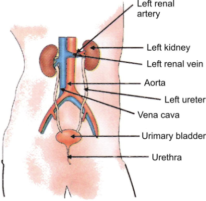
Human Excretory System: Human excretory system includes a pair of kidneys, a pair of ureters, a urinary bladder and a urethra. Urine produced in the kidneys passes through the ureters into the urinary bladder where it is stored until it is released through the urethra.
|
(a) Kidney
|
Excretory system in human beings |
Structure of Nephron
- Each kidney is made up of a large number of excretory filtration units called nephrons or uriniferous tubules.
- These are considered as functional unit of kidney.
- Each nephron has a cup shaped upper end, called Bowman’scapsule containing a bundle of capillaries called glomerulus. These blood capillaries arise from the artery, which brings waste and excess water from body to kidney. Bowman’s capsule leads into tubular structures. The tubular parts of many nephrons drain into a common collecting duct.
- The function of glomerulus is to filter the blood passing through it.
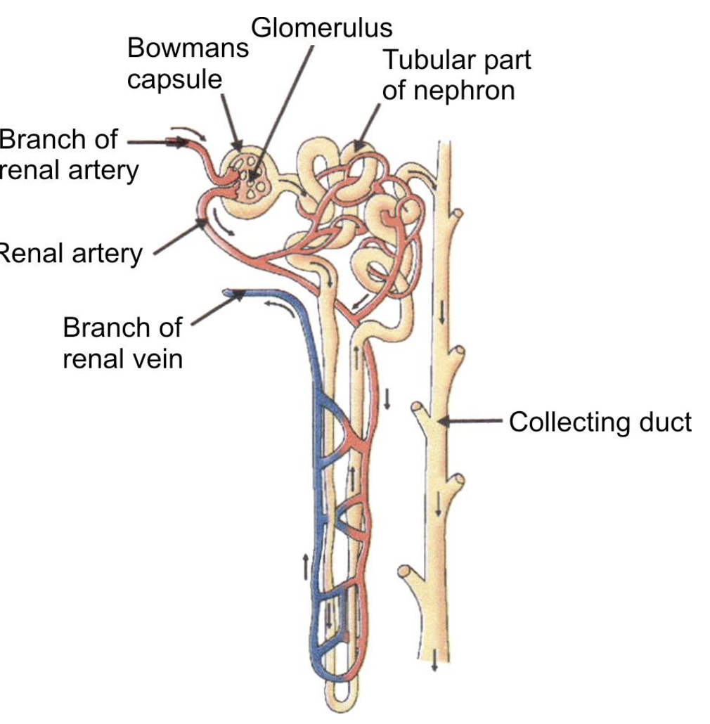
- The function of tubular part of nephron is to allow the selectivereabsorption of useful substances into the blood capillaries.
Blood having Metabolic Waste → Afferent Arteriole → Glomerulus → Bowman’s Capsule → PCT (Proximal Convoluted Tubule) → Loop of Henle → DCT (Distal Convoluted Tubule) → Collecting Duct → Ureter → Urinary bladder → Urethra → Urine excreted out
(b) Formation of urine: The purpose of excretion is to filter out waste products from the blood.
- The waste material is brought to kidneys by the renal arteries.
- Blood is filtered, from the blood capillaries into Bowman’s capsule.
- As this filtrate passes through the tubular parts of nephron, some useful products, such as glucose, amino acids, salts and major amount of water are selectively reabsorbed by blood capillaries surrounding the nephron.
- The nephrons drain the remaining liquid waste (urine) into the collecting duct which eventually enters a long tube, the ureter. Human urine contains water and nitrogenous substances, most of which is urea.
From the ureter, urine passes into the urinary bladder. Urine is stored in the urinary bladder until the pressure of the expanded bladder leads to the urge to pass it out through the urethra. The bladder is muscular, so it is under nervous control. As a result, we can usually control the urge to urinate.
(c) Artificial kidney: It is a device to remove nitrogenous waste products from the blood through dialysis. In case of kidney failure an artificial kidney can be used.
Dialysis: It is the procedure used in artificial kidney to do the work of a non-functional or damaged kidney. In the process, blood of the patient is allowed to pass through the long cellulose tubes dipped in a tank containing dialysing solution having same ionic concentration as plasma. The waste substance diffuse out of blood into the tank and the cleansed blood is returned back into the patient through a vein. This procedure is also known as haemodialysis.
SOLVED QUESTIONS
1. Name the process by which plants make food.
Sol. Photosynthesis
2. Where do plants get each of the raw materials required for photosynthesis?
Sol. CO2 from atmosphere and water from the soil.
3.(a) Name the gas used in photosynthesis.
(b) Name the gas produced in photosynthesis
Sol. (a) CO2 (Carbon dioxide)
(b) Oxygen (O2)
4. What kind of nutrition occurs in fungi?
Sol. Saprophytic
5. What are the end products of anaerobic respiration in yeast?
Sol. C2H5OH
6. Name one substance which is produced in anaerobic respiration but not in aerobic respiration.
Sol. Ethanol (Ethyl alcohol) and Lactic acid.
7. What is the medium of exchange of gases in aquatic animals?
Sol. Water
8. Name the site of aerobic respiration in cells?
Sol. Mitochondria
9. Why is anaerobic respiration considered less efficient?
Sol. It is a process of incomplete oxidation of glucose and it also releases less energy (2 ATP) as compared to aerobic respiration (38 ATP).
10. What is the full form of ATP?
Sol. Adenosine tri-phosphate.
11. Name the fluids that carry various substances in our body?
Sol. Lymph and blood
12. Name the two parts of vascular system in plants.
Sol. Xylem and phloem.
13. How is food transported in plants?
Sol. Food is transported through phloem in the dissolved form.
14. Name the excretory unit of a kidney.
Sol. Nephron
15. Name two functions performed by kidneys.
Sol. (a) Osmoregulation
(b) Excretion of urine
16. Why is it necessary to separate oxygenated and deoxygenated blood in mammals and birds?
Sol. Mammals and birds are warm blooded animals. To maintain their body temperature hey need energy which is obtained by aerobic respiration. Separation of oxygenated and deoxygenated blood is necessary for highly efficient supply of oxygen to the body cells for aerobic respiration so that energy requirement can be accomplished.
17. What are the components of the transport system in highly organized plants?
Sol. The transport system in highly organized plants consists of –
(a) Xylem – Transports water. Consists of tracheids and vessels
(b) Phloem – Transports food. Consists of sieve tubes and companion cells.
18. How are water and minerals transported in plants?
Sol. Water and minerals are transported through xylem, which consists of xylem vessels and tracheids forming a continuous water conducting channel which reaches all parts of the plants. Water and minerals are absorbed by the xylem of roots through osmosis and because of transpirational pull water moves in upward direction.
19. How is oxygen and carbon dioxide transported in human beings?
Sol. Oxygen is transported in combined form with respiratory pigment, Haemoglobin present in RBCs. Carbon dioxide is more soluble in water as compared to oxygen, so it is transported mainly in dissolved form and partly with haemoglobin in our blood.
20. What is the function of ATP?
Sol. ATP is the energy carrier molecule therefore it is used in body for muscle contraction, protein synthesis, conduction of nerve impulses and all other metabolic activities.
21. How aquatic animals respire?
Sol. Aquatic animals use the O2 dissolved in water. Since the amount of O2 in water is fairly low as compared to the amount of O2 in the air, the rate of breathing in aquatic organisms is much faster than that seen in terrestrial organisms.
22. How are the lungs designed in human beings to maximize the area for exchange of gases?
Sol. The trachea divides into bronchi, which enters the lungs. In the lungs bronchi divides into bronchioles which finally terminate into balloon like structures called alveoli. These alveoli provide a large surface area where the exchange of gases can take place. The wall of alveoli contains a fine network of blood capillaries for easy exchange of gases.
23. How does diaphragm help in respiration?
Sol. Contraction of diaphragm muscles causes flattening of diaphragm. This increases the volume of chest cavity, which creates low pressure in the lungs. As a result air is inhaled into the lungs. During exhalation, diaphragm relaxes and becomes arched. This decreases the volume of chest cavity increasing the air pressure and causing exhalation.
24. Draw a well labeled diagram of human respiratory system.
Sol.
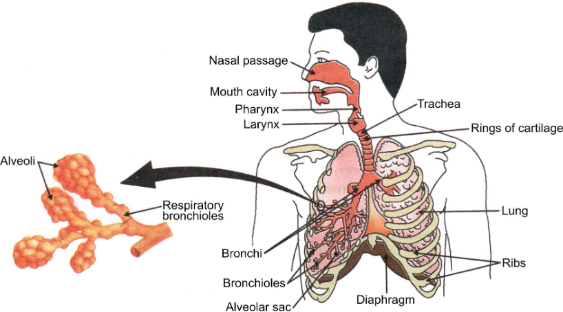
25. What are the methods used by plants to get rid of excretory products?
Sol. Plants use various methods to get rid of excretory products.
(a) By diffusion. O2 and CO2 diffuses in and out of stomata
(b) Transpiration. Excess water is removed by transpiration
(c) Some waste products are stored in cellular vacuoles.
(d) Resins and gums are stored in old xylem tissue.
(e) Plants also release some waste products into the soil around them.
(f) Some plant wastes are stored in leaves, which get removed when leaves fall off.