Sexual reproduction in flowering plants involves the formation of male and female gametes, their transfer, fusion, and development into seeds and fruits. The process begins with the flower, the reproductive organ of angiosperms. The male reproductive part, the stamen, produces pollen grains in the anthers, while the female part, the carpel (or pistil), contains the ovary with ovules. Pollination, the transfer of pollen from anther to stigma, may occur via wind, water, insects, or other agents. After pollination, the pollen grain germinates on the stigma and grows a pollen tube through the style to reach the ovule. Fertilization involves the fusion of one male gamete with the egg cell (syngamy) and another with the secondary nucleus (triple fusion), a process called double fertilization—unique to angiosperms. The fertilized ovule develops into a seed, and the ovary matures into a fruit. This method ensures genetic variation and plays a key role in the survival and adaptation of plant species.
Self pollination: This process involves the transfer of pollen grains from the anthers to the stigma of the same flower.
Contrivances or adaptations for self pollination
Homogamy: In some plants stamens and carpels of a flower mature at the same time. Therefore, pollen grains of a flower can pollinate the stigma of the same flower. Homogamy occurs in Mirabilis, Catheranthus roseus, Gardenia, potato, wheat, rice, etc.
Cleistogamy: Generally the flowers open after their maturation (anthesis) and expose their anthers and stigma to the environment. Such flowers are known as chasmogamous flowers. However, some flowers do not open even at maturity and are called cleistogamous flowers. Such cleistogamous flowers are forced to undergo self pollination. Cleistogamy is shown by the underground flowers of Commelina benghalensis (kanchara), Viola mirabilis, etc. Commelina also bears normal flowers on their aerial parts in addition to underground cleistogamous flowers.
Cross pollination:
- Agents and types of cross pollination. Cross pollination involves external agents for the transfer of pollen grains of one flower to the stigma of another flower
- Biotic agents which include animals of different types such as insects, birds, bats, snails, etc., and
- abiotic agents like wind and water.

Entomophily: Pollination by insects is known as entomophily. The common groups of insects bringing about pollination include bees, flies, wasps, drones, moths, butterflies, etc. About 80% of insect pollination is due to bees.
The insect pollinated flowers develop certain adaptations to attract them:
- Large sized flowers
- Bright coloured petals and perianth members
- Scent and odours
- Nectar in special glands
- Edible sap
- edible pollen
- special mechanisms
Special mechanism: Salvia often shows Lever or Piston mechanism of pollination. The flower has a bilabiate corolla with two epipetalous stamens. The two anther lobes of a stamen are widely separated by a long curved and lever-like connective. This connective moves freely on the filament. The upper anther lobe is fertile while the lower is sterile. Bees push against the lower anther lobe and swing the connective while entering the corolla tube in search of nectary situated at its base. This brings down the upper anther lobe which strikes the back of the insect and dusts it with pollen grains.
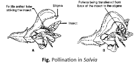
Ornithophily: There are only a few bird pollinated flowers. Humming bird and the honey thrushes feed on the nectar of flowers of Bignonia capreolata. Large flowers of Strelitzia raginae are pollinated by sun bird while collecting honey. The plants like Bombax (silk-cotton tree; semal), Erythrina (coral tree), Callistemon (bottle brush), etc., commonly found in the country, are pollinated by crows and mynas.
Cheiropterophily: This is the pollination by bats and commonly occurs in plants growing in tropical regions. Cheiropterophilous flowers are large sized, have long pedicels and produce large amount of nectar. Anthocephalus cadamba (Kadam), Kigelia africana (Sausage tree;), etc., are some of the common bat-pollinated plants.
Malacophily: In these cases pollinating agents are snails and slugs. Land plants like Chrysanthemum leucanthemum and water plant like Lemna show malacophily.
Anemophily: Wind pollinated or anemophilous flowers are small and inconspicuous. The pollen grains are often dry, small and light in weight. In some cases pollen grains become winged. The style and stigma are generally branched to catch the pollen grains floating in the air. In cereals. (e.g., maize) the styles are feathery appearing as tufts of fine, long and silky threads e.g. Gymnosperms, Grasses, Date palm, coconut etc.
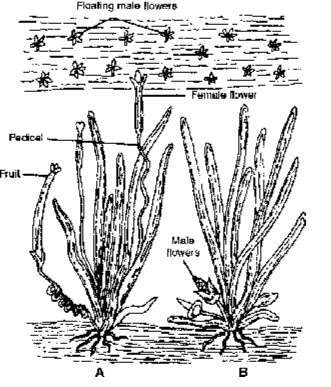
Hydrophily: The aquatic plants depend upon water for transfer of pollen grains; e.g., in plants belonging to families Najadaceae, Ceratophyllaceae, Potamogetonaceae, Hydrocharitaceae, etc. Pollination may take place in flowers occuring below the water level (Hypohydrophily) e.g. Ceratophyllum, Najas, Zostera etc, or in flowers floating on the water sufrace (Epihydrophily) e.g. Vallisneria, Hydrilla, Elodea etc.,
Contrivances (adaptations) for cross pollination
Dicliny or Unisexuality: These flowers are unisexual, i.e. male flowers possess only the stamens and female flowers possess carpels only.
Dichogamy: This is the condition where stamens and carpels of a bisexual flower mature at different times. Hence, self pollination between them remains ineffective. There are two different conditions
Protandry: In such flowers anthers mature earlier than the stigma of the flower. Protandry is commonly observed in cotton, lady’s finger, coriander, sunflower, etc.
Protogyny: In these flowers carpels mature earlier than the anthers of the flower. The common examples include banyan, peepal, custard apple, Magnolia, Michelia, etc.
Herkogamy: In some flowers, there is a physical barrier between anther and the style. This prevents self pollination. In members of Cruciferae and Caryophyllaceae, style is long and carries stigma far beyond the stamens. Such a stigma would not receive pollen from the same flower. In Calotropis and some orchids, pollens occur in special structures called pollinia (Translator). These can be transferred only with the help of insects.
Heterostyly: In some plants the flowers are of two (dimorphic) or three (trimorphic) forms with anthers and stigmas at different levels. Based on difference in length, the phenomenon is called heterostyly (styles of different lengths) and heteroanthy (stamens of different lengths). Pollination is convenient between stamens and styles of the same length borne by two different flowers. In such flowers there is also a possibility of self-pollination if cross pollination fails. Dimorphic heterostyly is shown by Primula (primrose), Fagopyrum, Linum (linseed), Oxalis (wood-sorrel), etc. Oxalis, Linum and Lythrum show trimorphic heterostyly.
Self sterility Pollen grains fail to germinate on stigma of the same flower. It is also known as intraspecific incompatibility. It is of two types:
- Sporophytic incompatibility: It is due to genotype of the sporophytic (stigmatic) tissues.
- Gametophytic incompatibility: It is due to genotype of pollen.
Post-fertilization Changes
Following are post-fertilization changes-endosperm formation, embryo formation, seed formation and fruit formation.
Endosperm (3N) Formation
Endosperm is meant for nourishing the embryo. In Angiosperms, it is triploid because it is formed after vegetative fertilization, where triple fusion takes place. Endosperm may show the effect of genes present in the male gamete.
Types of Endosperms
Nuclear Endosperm: Primary endosperm nucleus divides and redivides to form a large number of free nuclei. A central vacuole appears and a massive peripheral multinucleate cytoplasm is formed. The central vacuole disappears in Capsella bursa-pastoris. Cell wall formation is incomplete in Coconut where there is an outer multicellular solid endosperm and inner free nuclear liquid endosperm in the centre.
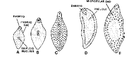
Cellular Endosperm: Wall formation occurs after every division of primary endosperm nucleus, so that endosperm is of cellular form from the beginning e.g., Datura, Petunia.
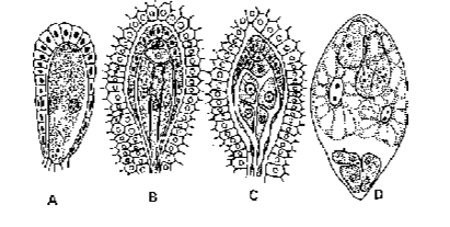
Helobial Endosperm: First division of primary endosperm nucleus is followed by cytokinesis to produce two cells; micropylar and chalazal within which free nuclear divisions occurs and cytokinesis takes place at the last step to make them cellular. e.g., Asphodelus.
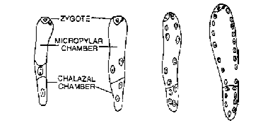
Endosperm may persist in the seed. It is called as endospermic seed or albuminous seed e.g.,Castor. It may be consumed by the embryo. In such a case food is generally stored in the cotyledons. Such seeds are called non-endospermic or exalbuminuous., Groundnut, Ruminate or convoluted endosperm occurs in Areca (Betelnut) and Passiflora. Hard endosperm is found in Date, Phytelepas and Areca.
Embryo Formation (Embryogeny)
It is the development of mature embryo from zygote or oospore. Development of embryo is endoscopic or on inner side due to presence of suspensor.
Embryonic Development in Dicotyledons
Embryonic Development in Dicotyledons(Crucifer/Onagrad type). Zygote undergoes elongation and divides transversely into two unequal cells i.e., terminal cell and suspensor cell.
Suspensor Cell, It is larger basal cell which undergoes transverse divisions to form 6 to 10 celled suspensor. First cell of the suspensor (toward micropyle) is large and called haustorium. Increase in number of suspensor cells pushes the terminal embryo cell into endosperm. The last cell of suspensor toward embryo cell is known as hypophysis. It later on results into the formation of radicle.
Embryo Cell (Terminal Cell) : It is present towards the antipodal side. It divides twice vertically and once transversely to produce a two-tiered 8-celled embryo.
- Epibasal (Terminal) Tier : It forms two cotyledons and a plumule. Later on, plumule gives rise to stem.
- Hypobasal Tier (Near Suspensor). It produces only hypocotyl. This octant embryo undergoes periclinal division producing two layers of cells.
Outer Layer. It will form protoderm or dermatogen for the formation of epidermis.
Inner Layer. It will form procambium (for the formation of vascular strand i.e., stele) and ground meristem (for the formation of cortex and pith)
Embryo is initially globular, undifferentiated and radially symmetrical. It is named as proembryo. Later on it develops into embryo with the formation of radicle, plumule and cotyledons. With the growth of cotyledons, It becomes heart shaped and then assumes, the typical shape. e.g.,m Capsella bursa-pastoris.
Embryonic Development in Monocotyledons (Sagittaria type) : The zygote divides transversely, producing a vesicular suspensor cell towards micropylar end and embryo cell towards the chalazal end. The embryo cell divides transversely again into a terminal cell and a middle cell. The terminal cell divides vertically and transversely into globular embryo. It forms a massive cotyledon and plumule. Growth of cotyledon pushes the plumule to one side.
Remains of second cotyledon occur in some grasses. It is called epiblast. The single cotyledon of monocots is called scutellum. It is shield shaped and appears terminal. The middle cell gives rise to hypocotyl and radicle. It may add a few cells to the suspensor. Both radicle and plumule develop covering sheaths called coleorhiza and coleoptile respectively. They appear to be extension of scutellum.
SEED
Morphologically, ripened ovule is known as seed. Seed can also be defined as a mature integumented megasporangium. It is a unit containing an embryo plant in which the primordia of future structures are already established and embedded in nutrient and protective tissue derived from previous generation.
Seed Coat: It is protective covering of the seed and is made up of two layers : (a) outer, called Testa, Which is usually hard, and (b) inner, called Tegmen, which is thin and papery
There is a small opening at one end of the seed coat called micropyle through which water enters. There is a depression like concavity at middle of one edge, where the seed breaks from the stalk of funiculus; this is called hilum. There is a ridge beyond the hilum opposite the micropyle. It represents the base of the funiculus which is fused with the integuments and is called raphe
Embryo: Embryo is a young plant enclosed in a seed coat. It has two parts :
- Cotyledons: These are leaves of embryo. Their number is either one or two. Sometimes they store food materials and become fleshy. When they do not store food they remain thin and papery. The cotyledons are hinged to an axis (tigellum) at a point called cotyledonary node and open out like a book.
- Tigellum: It is the main axis of the embryo. One end of the tigellum is pointed and protrudes out of cotyledons. This lies next to mircopyle and is called radicle (rudimentary root). The other end of tigellum is the feathery plumule ( first apical bud of shoot). The portion of axis lying between the cotyledonary node and plumule is called epicotyl while the portion between the node and radicle is called hypocotyl.
Endosperm: It is food-laden tissue, either present on one side of embryo or surrounding the embryo on all sides. In some seeds, the endosperm is present until maturity. Such seeds are called endospermic (albuminous) seeds. In some seeds it is consumed in young stages by the delveloping cotyledons and such seeds do not have endosperm at maturity. Such seeds are called non endospermic(ex-albuminous) seeds.
All parts enclosed by seed coat are called kernel.
Pistil, Megasporangium (ovule) and embryo sac
The gynoecium represents the female reproductive part of the flower. The gynoecium may consist of a single pistil (monocarpellary) or may have more than one pistil (multicarpellary). When there are more than one, the pistils may be fused together (syncarpous) or may be free (apocarpous). Each pistil has three parts, the stigma, style and ovary. The stigma serves as a landing platform for pollen grains. The style is the elongated slender part beneath the stigma. The basal bulged part of the pistil is the ovary. Inside the ovary is the ovarian cavity (locule). The placenta is located inside the ovarian cavity. Arising from the placenta are the megasporangia, commonly called ovules. The number of ovules in an ovary may be one (wheat, paddy, mango) to many (papaya, water melon, orchids).
Ovule, Megasporogenesis and the Megaspore
The ovule: Ovule is considered to be an integumented megasporangium. The ovules are situated inside the ovary, attached to a special tissue called placenta.
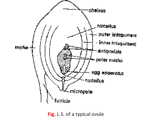
Structure of ovule
The stalk is called funicle. One end of the funicle is attached to placenta and the other end to the body of the ovule. The point of attachment of funicle with the body is called hilum. Funicle sometimes extends up to the base of ovule (i.e. chalaza). The ridge thus formed is called raphe.
The body of the ovule shows two ends- the basal end, often called the chalaza and the upper end is called the micropylar end. The main body of the ovule is covered with one or two envelops called integuments. These leave an opening at the top of the ovule called micropyle. The integuments enclose a large parenchymatous tissue known as nucellus. In the centre of the nucellus is situated a female gametophyte known as embryosac.
Following are some of the conditions seen in ovule in relation to integuments.
- Unitegmic Ovule with a single integument; e.g. gamopetalous dicotyledons.
- Bitegmic Ovule with two integuments as in polypetalous dicotyledons and monocotyledons.
- Aril This is a collar-like outgrowth from the base of the ovule and forms third integument. Aril is found in litchi, nutmeg, etc.
- Caruncle It is formed as an outgrowth of the outer integument in the micropylar region. Caruncle is common in the ovules of Euphorbiaceae e.g. castor.
- Ategmic In some parasites like Loranthus, Viscum, Santalum, etc. there is no integument, such an ovule is called ategmic.
Types of ovules
There are six types of ovules in Angiosperms. These are classified on the basis of mutual relationship between funicle, chalaza and micropyle.
- Atropous or orthotropous: In this type, funicle, chalaza and micropyle are situated on one vertical axis with micropyle directed upwards. Hence it is also known as straight ovule; e.g. Polygonum, Piper etc.
- Anatropous: These ovules are also called inverted (180° curved) because the body of the ovule is so bent that micropyle is now directed downwards and lies close to the hilum. It is the most common type of ovule found in Angiosperms, e.g., castor.

- Campylotropous: This is an ovule placed transversely on the funicle (90° curved). The body is bent in such a way that micropyle gets directed downwards and is not in straight line with chalaza. It is found in gram, bean, members of Cruciferae like mustard etc.
- Hemianatropous:In this type, the body of the ovule is situated at right angles to the funicle e.g. Ranunculus.
- Amphitropous:The ovule is curved (at 180°) and embryo sac also becomes horse shoe-shaped. The micropyle is directed downwards and does not lie on straight line with chalaza. Amphitropous ovules occur in poppy, members of Alismaceae, Butomaceae, etc.
- Circinotropous:The ovule is initially orthotropous but becomes anatropous due to unilateral growth of funicle. The growth continues till the ovule once again becomes orthotropous. As a result funicle completely surrounds the body of the ovule; e.g. Opuntia (prickly pear).
Megasporogenesis
The process begins with the differentiation of archesporial initial in the nucellar hypodermis. Archesporial initial either acts directly as a megaspore mother cell or divides periclinally into an outer primary parietal cell and the inner primary sporogenous cell. Accordingly, following two conditions are found.
Tenuinucellate ovule: In this condition, the archesporial initial directly behaves as megaspore mother cell. Hence in this case primary parietal cell is not formed. These ovules are called tenuinucellate and are found in gamopetalous dicotyledons with unitegmic ovules.
Crassinucellate ovule: The division of the archesporial initial into primary parietal cell and primary sporogenous cell results in the formation of crassinucellate ovule. The primary parietal cell, thus formed, divides to give rise to a large amount of nucellar tissue between primary sporogenous cell and nucellar epidermis. Such ovules occur in polypetalous dicotyledons with bitegmic ovules.
The megaspore mother cell (megasporocyte) divides meiotically to form a linear tetrad of four haploid megaspores. Occasionally, T-shaped or inverted T-shaped () tetrads are also formed. Megaspore is the first cell of female gametophyte.
Of the linear tetrad, three megaspores towards the micropyle degenerate. The lowermost, i.e. the chalazal megaspore enlarges and remains functional. It later produces an embryo sac.
Development of Embryo sac (Female Gametophyte)
On the basis of their development, following three types of embryo sacs can be distinguished. These are monosporic, bisporic and tetrasporic.
Monosporic embryo sac: In this type only one out of the four megaspores takes part in the development of embryo sac. A monosporic 8-nucleate embryo sac, known as Polygonumtype, occurs in about 70% of Angiosperms and is the most common type. Oenothera is a monosporic but four nucleate embryo sac.
During the development of Polygonum type of embryo sac, functional megaspore undergoes three mitotic divisions to form 8-nucleate embryo sac. Of these, four nuclei occur at micropylar end and the other four at the chalazal end.
- Three nuclei at the micropylar end form egg apparatus and the fourth migrates to the centre and forms one of the two polar nuclei.
- At the chalazal end, three nuclei form antipodal cells and the remaining fourth migrates to the centre to form second polar nucleus.
A fully developed monosporic 8-nucleate polygonum type of embryo sac shows following organisation:
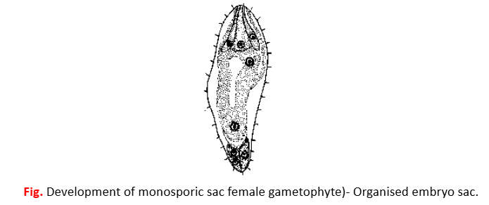
- Egg apparatus This is a group of 3 cells situated at the micropylar end. The centrally located cell is called egg cell. On its sides are present two synergids. Egg cell has a large vacuole at its upper end and a prominent nucleus near its lower end. Synergids show a filiform apparatus attached to their upper wall. It is known to attract and guide the pollen tube. Each of the synergids has a vacuole at its lower end and the nucleus at its upper end.
- Polar nuclei Generally, both the polar nuclei fuse before fertilization and form a single diploid nucleus called secondary nucleus or definitive nucleus.
- Antipodals The three cells situated at the chalazal end are called antipodals. These cells generally degenerate soon after fertilization.
In some members of Gentiniaceae,the three antipodal cells divide to form about ten to twelve cells,and in the family Graminae a still larger number of cells is produced.
Thus a mature monosporic Polygonum type of embryo sac at the time of fertilization is 7-nucleate.
Bisporic embryo sac: In this type two megaspore nuclei take part in embryo sac formation.
e.g. Allium, Endymion.
Tetrasporic embryo sac: This embryo sac develops from four megaspore nuclei.
Frequently Asked Questions
The flower is the reproductive organ, containing male (stamens) and female (carpels/pistils) structures.
It is the process of formation of microspores (pollen grains) from pollen mother cells (PMCs) in the anther by meiosis.
It is the formation of megaspore from the megaspore mother cell (MMC) in the ovule through meiosis.
Pollination is the transfer of pollen grains from the anther to the stigma. It can be self-pollination (autogamy, geitonogamy) or cross-pollination (xenogamy).
Wind (anemophily), water (hydrophily), insects (entomophily), birds (ornithophily), bats (chiropterophily), etc.
In flowering plants, one male gamete fuses with the egg cell (syngamy) to form the zygote, and the other fuses with the secondary nucleus (triple fusion) to form the endosperm. This is double fertilization and is unique to angiosperms.
The endosperm provides nutrition (starch, oils, proteins) to the developing embryo.
The fertilized ovule develops into a seed, consisting of an embryo, stored food, and a seed coat.
- Albuminous: Endosperm persists (e.g., wheat, maize).
- Exalbuminous: Endosperm is used up during seed development (e.g., pea, groundnut).
It is the formation of seeds without fertilization, producing genetically identical offspring.