Animal Tissues: A Comprehensive Study Guide
Animal tissues form the fundamental building blocks of multicellular organisms, representing groups of similar cells working together to perform specific functions. This comprehensive guide explores the four primary types of animal tissues, their characteristics, functions, and biological significance.
What Are Animal Tissues?
A tissue is defined as a group of cells that share similar structure, origin, and function. The term "tissue" was coined by French anatomist and physiologist Bichat (1771-1802), while the study of tissues, known as histology, was named by German histologist Mayer in 1819. Italian scientist Marcello Malpighi (1628-1694) is recognized as the "founder of histology."
Key Terminology in Tissue Biology
- Histology: The microscopic study of tissues (also called microanatomy)
- Histogenesis: Formation and development of tissues from undifferentiated embryonic cells
- Histodifferentiation: The process by which cell groups differentiate into tissues
- Histolysis: Degeneration of tissues
Classification of Animal Tissues
Animal tissues are classified into four fundamental types based on their location, structure, and function:
- Epithelial Tissue
- Connective Tissue
- Muscular Tissue
- Nervous Tissue
Epithelial Tissue: The Protective Barrier
Definition and Characteristics
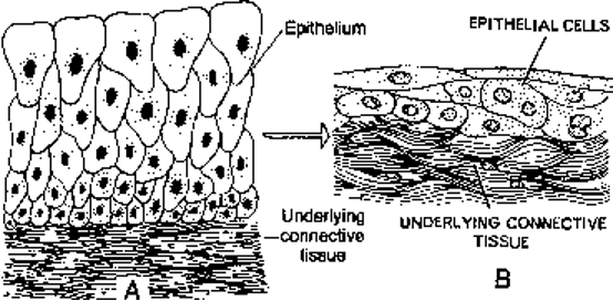
Epithelial tissue consists of one or more layers of compactly arranged cells overlying a non-cellular basement membrane. The term "epithelium" was coined by Dutch anatomist Ruysch (1638-1731), meaning "tissues that grow upon other tissues."
Key Features of Epithelial Tissue
- Cellular arrangement: Cells are closely packed with minimal intercellular space
- Basement membrane: Non-cellular layer beneath the epithelium with two parts:
- Basal lamina (outer layer)
- Reticular lamina (inner layer)
- Avascular nature: No blood vessels; nutrition provided by diffusion
- Regenerative capacity: Rapid cell division and replacement
- Cell shape: Typically polygonal, varying based on function
Types of Epithelial Tissue
A. Covering and Lining Epithelium
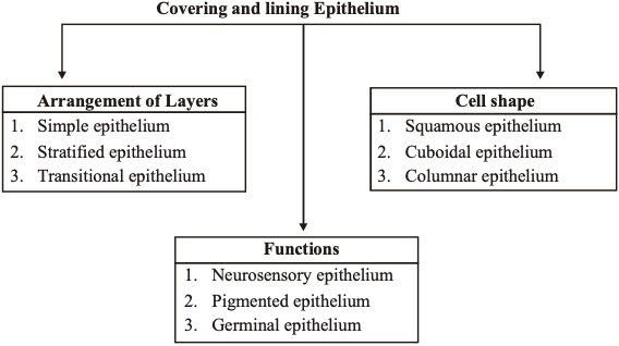
1. Simple Epithelium (Single Layer)
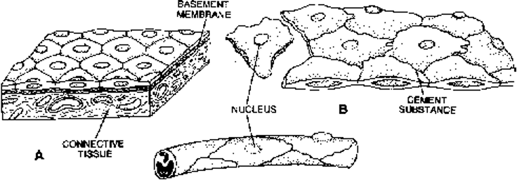
Simple Squamous Epithelium
- Structure: Flat, tile-like polygonal cells with centrally located nuclei
- Alternative name: Pavement epithelium
- Location: Terminal bronchioles, alveoli of lungs, Bowman's capsule, loop of Henle
- Function: Protection, gas exchange, secretion of coelomic fluid
Simple Cuboidal Epithelium
- Structure: Single layer of cube-shaped cells with spherical nuclei
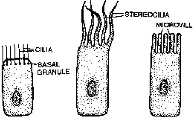
- Special feature: May form microvilli (brush-bordered cuboidal epithelium)
- Location: Thyroid vesicles, kidney tubules, ovaries, seminiferous tubules
- Function: Protection, secretion, absorption, excretion, gamete formation
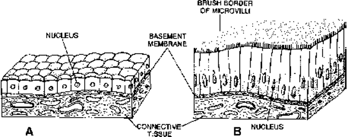
Simple Columnar Epithelium
- Structure: Tall, column-like cells with elongated nuclei in basal region
- Special cells: Contains goblet cells that secrete mucus
- Location: Stomach, small and large intestine, digestive glands
- Function: Absorption, secretion, protection
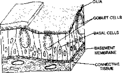
Simple Ciliated Epithelium
- Structure: Cells bear hair-like projections (cilia) from basal granules
- Types:
- Ciliated columnar (respiratory tract, fallopian tubes)
- Ciliated cuboidal (kidney nephrons)
- Function: Movement of materials in one direction
Pseudostratified Epithelium
- Structure: Appears multi-layered due to nuclei at different levels
- Reality: Single cell layer with cells of unequal height
- Location: Trachea, large bronchi, male urethra
- Function: Protection, secretion, movement of materials
2. Stratified Epithelium (Multiple Layers)
Stratified Squamous Epithelium
- Types:
- Keratinized: Contains waterproof keratin protein (skin epidermis)
- Non-keratinized: Lacks keratin (oral cavity, esophagus, vagina)
- Layers:
- Stratum germinativum (basal layer)
- Intermediate layers
- Squamous layers (superficial)
Transitional Epithelium (Urothelium)
- Structure: 4-6 layers with umbrella-shaped surface cells
- Location: Urinary bladder, ureters, renal pelvis
- Function: Permits distention without damage
B. Glandular Epithelium
Specialized epithelial cells forming glands can be classified based on:
1. Nature of Secretion
- Mucous glands: Secrete slimy mucin (goblet cells)
- Serous glands: Produce clear, watery secretions (parotid gland)
- Mixed glands: Both mucous and serous components (submandibular gland)
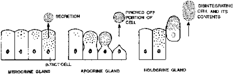
2. Mode of Secretion
- Merocrine: Cells remain intact during secretion (most sweat glands)
- Apocrine: Part of cytoplasm pinched off (some sweat glands)
- Holocrine: Entire cell disintegrates (sebaceous glands)
3. Duct System
- Exocrine: Secretions released through ducts (salivary glands)
- Endocrine: Ductless glands secreting into bloodstream (thyroid)
- Paracrine: Local hormone action (prostaglandins)
Connective Tissue: The Body's Framework
Origin and General Characteristics
Connective tissue originates from the embryonic mesoderm and represents the most abundant tissue type, comprising about 30% of body mass. It consists of fewer cells, extensive extracellular matrix, and rich blood supply.
Components of Connective Tissue
1. Intercellular Matrix (Ground Substance)
- Primarily composed of hyaluronic acid and other mucopolysaccharides
- Provides medium for nutrient and waste transport
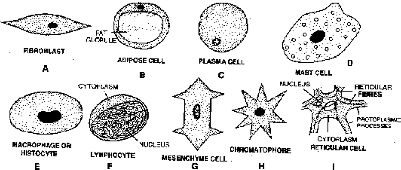
2. Connective Tissue Cells
Fibroblasts
- Function: Produce fibers and matrix components
- Most abundant: Found throughout connective tissues
Adipose Cells (Adipocytes)
- Function: Store fat for energy and insulation
- Types: White fat (single large droplet) and brown fat (multiple small droplets)
Plasma Cells
- Function: Synthesize antibodies
- Alternative name: "Cart wheel cells" due to nuclear appearance
Mast Cells
- Function: Produce histamine, heparin, and serotonin
- Role: Inflammatory and allergic reactions
Macrophages
- Function: Phagocytosis of debris and pathogens
- Origin: Derived from blood monocytes
3. Connective Tissue Fibers
| Fiber Type | Protein | Characteristics | Function |
|---|---|---|---|
| Collagen (White) | Collagen | Unbranched, inelastic, bundled | Tensile strength |
| Elastic (Yellow) | Elastin | Branched, elastic | Flexibility and stretch |
| Reticular | Reticulin | Delicate, branched, network-forming | Support framework |
Types of Connective Tissue
1. Connective Tissue Proper
Areolar Tissue (Loose Connective Tissue)
- Most widely distributed connective tissue
- Components: All fiber types, various cell types in ground substance
- Location: Subcutaneous layer, around organs
- Functions: Binding, support, rapid diffusion, immune cell migration
Adipose Tissue
- Specialized loose connective tissue for fat storage
- Function: Energy storage, insulation, organ protection
- Types: White fat (energy storage) and brown fat (heat production)
Dense Connective Tissue
- White Fibrous: Predominantly collagen fibers
- Examples: Tendons (muscle to bone), ligaments (bone to bone)
- Yellow Elastic: Predominantly elastic fibers
- Examples: Arterial walls, vocal cords, ligamentum flavum
2. Skeletal Tissues
Cartilage
- Structure: Chondrocytes in lacunae within chondrin matrix
- Growth: Unidirectional from periphery
- Types:
- Hyaline: Most common, translucent matrix (nose, larynx, joints)
- Elastic: Contains elastic fibers (ear pinna, epiglottis)
- Fibrocartilage: Dense collagen bundles (intervertebral discs)
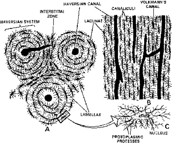
Bone
- Hardest tissue in the body
- Composition: 67% mineral salts (hydroxyapatites), 33% organic matrix (collagen)
- Structure: Osteocytes in lacunae, connected by canaliculi
- Haversian system: Characteristic of mammalian long bones
- Types: Compact (dense) and spongy (cancellous) bone
3. Vascular Tissues
Blood
- Composition: 55% plasma, 45% cellular components
- pH: 7.4 (slightly alkaline)
- Functions: Transport of gases, nutrients, wastes, hormones
Blood Components:
- Plasma proteins: Albumin, globulins, fibrinogen
- Erythrocytes (RBCs): Oxygen transport via hemoglobin
- Leukocytes (WBCs): Immune defense
- Platelets: Blood clotting
Lymph
- Colorless fluid without RBCs and platelets
- Composition: Plasma and lymphocytes
- Functions: Protein return, immune surveillance, fat transport
Muscular Tissue: The Movement Generator
General Characteristics
Muscular tissue originates from mesoderm and comprises about 40% of body weight in mammals. The specialized properties include contractility and electrical excitability.
Types of Muscular Tissue
1. Skeletal Muscle (Striated)
- Appearance: Cross-striations due to actin and myosin arrangement
- Control: Voluntary (somatic nervous system)
- Structure: Multinucleated syncytium
- Bands: A-bands (dark), I-bands (light), Z-lines, H-zones
- Contraction mechanism: Sliding filament theory
Muscle Fiber Organization:
- Endomysium: Surrounds individual fibers
- Perimysium: Surrounds fasciculi (bundles)
- Epimysium: Surrounds entire muscle
2. Smooth Muscle (Non-striated)
- Appearance: No cross-striations
- Control: Involuntary (autonomic nervous system)
- Structure: Spindle-shaped, uninucleated cells
- Location: Visceral organs, blood vessels
- Types: Single-unit and multi-unit smooth muscle
3. Cardiac Muscle
- Appearance: Striated but branched
- Control: Involuntary with intrinsic rhythm
- Structure: Uninucleated cells with intercalated discs
- Location: Heart wall and large veins
- Special features: Never fatigues, rich blood supply
Nervous Tissue: The Communication Network
Basic Components
Nervous tissue contains two main cell types:
- Neurons: Conducting cells
- Neuroglia: Supporting cells
Neuron Structure and Function
Neuron Components
Cell Body (Cyton/Soma)
- Contains nucleus and most organelles
- Nissl bodies: RNA-rich granules for protein synthesis
- Location of metabolic activities
Dendrites
- Function: Conduct impulses toward cell body
- Structure: Highly branched extensions
- Classification: Afferent processes
Axon
- Function: Conduct impulses away from cell body
- Structure: Single, long process with terminal branches
- Classification: Efferent process
- Covering: May be myelinated or non-myelinated
Types of Neurons
| Type | Processes | Location | Function |
|---|---|---|---|
| Unipolar | Single process | Embryonic tissue | Development |
| Bipolar | Two processes | Retina, inner ear | Sensory |
| Multipolar | Multiple dendrites, one axon | Brain, spinal cord | Integration |
Functional Classification
- Sensory neurons: Carry information to CNS
- Motor neurons: Carry commands from CNS
- Interneurons: Connect sensory and motor neurons
Neuroglia (Supporting Cells)
Types in CNS
- Astrocytes: Star-shaped, maintain blood-brain barrier
- Oligodendrocytes: Form myelin sheaths in CNS
- Microglia: Phagocytic cells, immune function
- Ependymal cells: Line ventricles, produce cerebrospinal fluid
Integration and Clinical Significance
Tissue Interactions
Animal tissues work together to form organs and organ systems. Understanding tissue types is crucial for:
- Medical diagnosis: Histopathological examination
- Surgical procedures: Knowledge of tissue properties
- Regenerative medicine: Tissue engineering applications
- Drug development: Tissue-specific drug delivery
Study Tips for CBSE Students
- Create comparison charts for different tissue types
- Practice labeling diagrams of tissue structures
- Memorize locations and functions of each tissue type
- Understand the relationship between structure and function
- Review clinical applications and examples
Important Examination Points
- Differences between tissue types (structure, function, location)
- Epithelial tissue classification and examples
- Connective tissue components and their roles
- Muscle contraction mechanism and types
- Neuron structure and signal transmission
- Tissue modifications and adaptations
This comprehensive guide provides the foundational knowledge necessary for understanding animal tissues in CBSE biology. Regular review and application of these concepts will ensure thorough preparation for examinations and future biological studies.