All living things, including animals, are made up of cells. In animals, similar cells join together to form a tissue. A tissue is a group of cells that have the same structure and work together to do a specific job. This organisation makes the animal’s body more efficient and helps it perform many functions at the same time.
In simple animals, the body may be made up of only a few types of cells. But in complex animals, there are many kinds of tissues, each with a special role. The main types of animal tissues are epithelial tissue, connective tissue, muscular tissue, and nervous tissue.
- Epithelial tissue covers the surface of the body and lines the organs. It protects the body and can also absorb, secrete, or sense things.
- Connective tissue supports and binds different parts of the body. Examples include bones, cartilage, blood, and fat tissue.
- Muscular tissue helps in movement. It can be voluntary (we control it) or involuntary (works automatically).
- Nervous tissue carries messages between the brain, spinal cord, and the rest of the body. It helps animals sense and respond to changes.
The way tissues are arranged in animals shows structural organisation – from cells to tissues, tissues to organs, and organs to organ systems. For example, muscle tissue in the heart works together with connective and nervous tissue to pump blood. Understanding animal tissues helps us know how the body works, how different systems are connected, and how injuries or diseases affect animals. It also helps in medical treatments, veterinary science, and biology research.
In short, tissues are the building blocks of animal bodies, making life possible by performing essential tasks every moment.
Tissues
A group of similar cells along with intercellular substances that usually have a common embryonic origin and function to carry out specialized activities. Body tissue can be classified into four principle types, according to their function and structure.
| Epithelial | Connective | Muscle | Nervous |
|
|
|
|
Epithelial Tissue and Its Classification
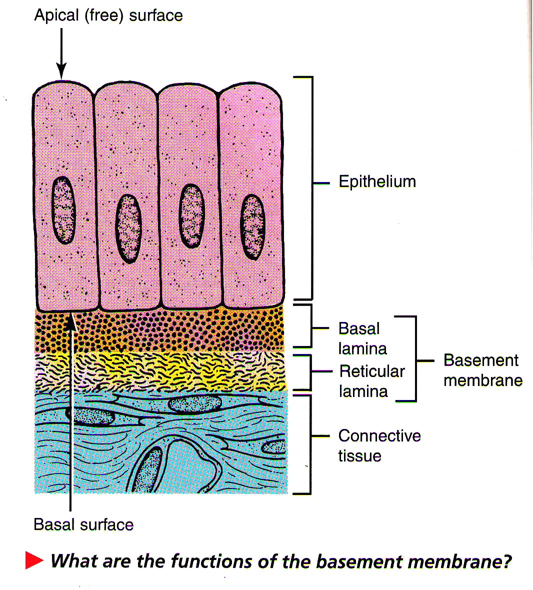
- Apical free surface which faces either body fluid or the outside environment.
- The cells are compactly packed with little intercellular matrix.
- Presence of basement membrane, except transitional epithelium.
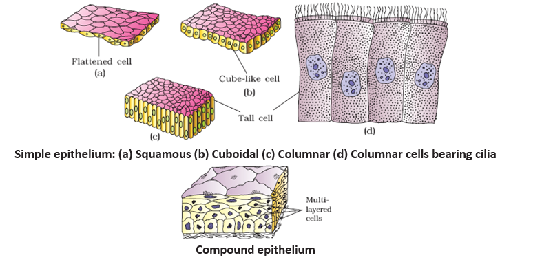
On the basis of cell layers and structural modification of the cells, epithelial tissue can be classified as:
| Simple | Compound | Pseudo Stratified |
| Composed of single layer of cells which serves for absorption or filtration. | Composed of more than one layer of cells. Limited role in secretion and absorption, its main function is to provide protection against chemical and mechanical stress. | Cells of different sizes are arranged as single layer. |
Classification on the basis of cell types, its arrangement and distribution
| Type | Function | Location |
| Simple squamous epithelium | Filtration, diffusion, osmosis | Air sacs of lungs, walls of capillaries, lining of blood and lymph vessels. |
| Simple cuboidal epithelium | Secretion, absorption | Surface of ovaries, lining of kidney tubules, and lining of ducts of certain glands |
| Simple columnar epithelium | Protection, secretion, absorption | Lining of uterus and organs of the digestive tract |
| Pseudostratified columnar epithelium | Protection, secretion, movement of mucus and cells | Lining of respiratory passages and reproductive tract |
| Stratified squamous epithelium | Protection | Outer layer of skin, lining of oral cavity, throat, vagina, and anal canal |
| Stratified cuboidal epithelium | Protection, secretion | Male urethra, parts of the pharynx |
| Transitional epithelium | Distensibility, protection | Inner lining of urinary bladder and passageways of urinary tract |
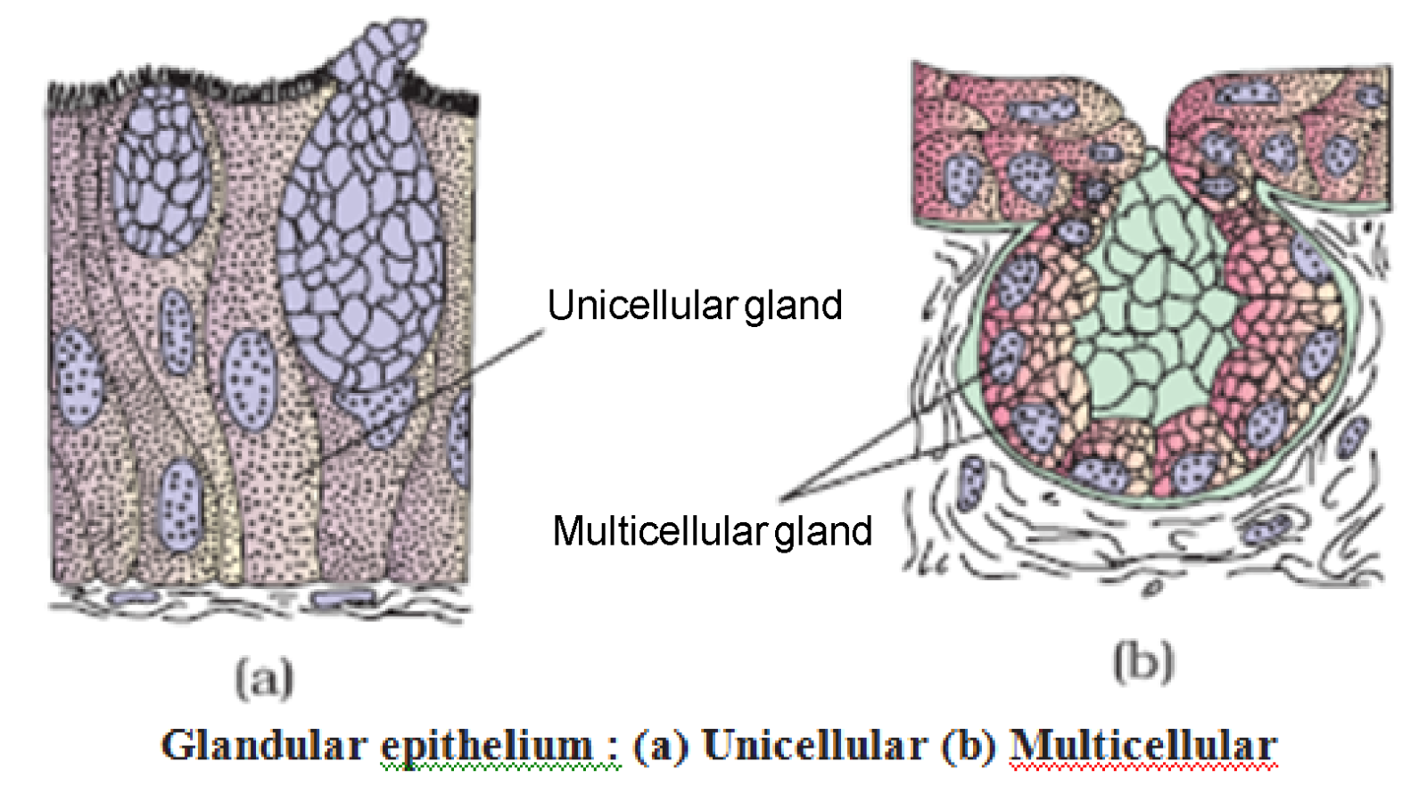
Glandular Epithelium
Composed of cells that are specialized to produce and secrete substances into ducts or into body fluids. Such cells are usually found in columnar or cuboidal epithelium.
Glands – Based on number of cells
| Unicellular | Multicellular |
| e.g., Goblet cells of alimentary canal. | e.g., Salivary glands, gastric gland, mammary gland. |
Glands – Based on secretion
| Exocrine Gland | Endocrine Gland |
| Secrete their products into ducts that open into some internal or external surface. e.g. – Gland which secrete mucus, saliva, earwax, oil, milk, digestive enzymes and other cell products. | Secrete their products into tissue fluid or blood. e.g. – Hormone secreting glands. |
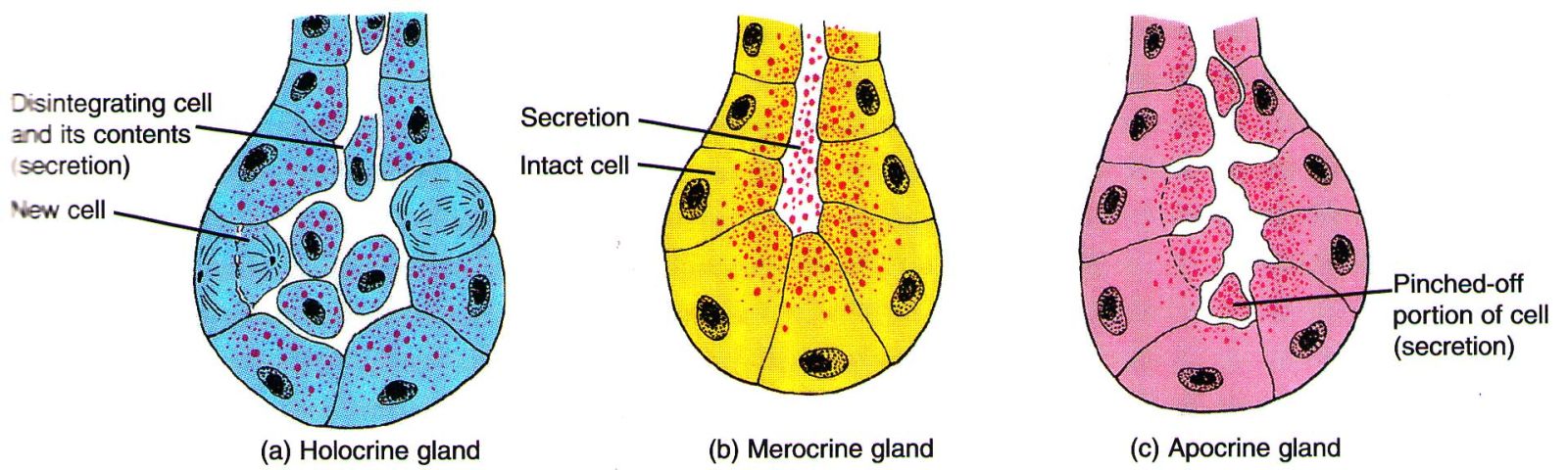
Functional Classification of Exocrine Gland
Holocrine Gland
An entire cell when filled with secretory products dies and disintegrates and is therefore discharged as a part of secretion (sebaceous gland).Merocrine or Epocrine Gland
Secretion is discharged by the cells by simple diffusion so that there is no loss of the cells (most common exocrine gland). e.g., goblet cell, sweat gland, gastric glandApocrine Gland
Secretory products accumulate in the apical parts of the cell; later this part breaks off from remaining part of a cell and is discharged as secretion. e.g., mammary gland
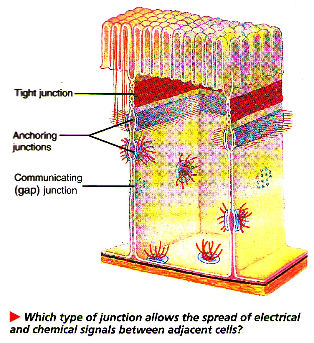
Cell Junction
Most epithelial cells, some muscle cells and some nerve cells are tightly joined to form a close functional unit. The points of contact between adjacent plasma membranes are called cell junctions.
| Tight Junction | Adhering or Anchoring Junction | Gap Junction |
| It forms fluid tight seals between cells which prevents substances from leaking across a tissue. | It performs cementing to keep neighbouring cells together and is common in tissues subjected to friction and stretching. | It provides cytoplasmic connection and allows electrical or chemical signals to pass from cell to cell. |
| Present between the cells of stomach, intestine and urinary bladder. | Outer layer of skin, muscle tissue of the heart. | Present in nervous tissue, muscle of the heart and gastrointestinal tract. |
Connective Tissue
This tissue provides the structural framework and support to different tissues, helps in body defense, tissue repair and fat storage etc. About 30% of the body mass is formed of connective tissue. It is most abundant and most widely distributed tissue in the body.
Components of Connective Tissue
It consists of various types of cells along with ground substance or matrix. It mainly consists of water and sulfated mucopolysaccharides.
| Component | Characteristic | Function |
| Fibroblasts | Widely distributed, large, star-shaped cells | Secrete proteins that becomes fibres |
| Macrophages | Motile cells that are sometimes attached to fibres | Engulf foreign particles from tissues by phagocytosis |
| Mast cells | Large cells, usually located near blood vessels | Releases substances that may help prevent blood clotting and promote inflammation |
| Collagenous fibres (white fibres) | Thick, thread like fibres of collagen with great tensile strength | Hold structures together |
| Elastic fibres (yellow fibres) | Bundles of microfibrils composed of elastin that is very elastic | Provide elastic quality to parts that stretch |
| Reticular fibres | Thin fibres of collagen | Form supportive network within tissues |
Classification of Connective Tissue (CT)
| CT Proper | Supporting CT | Fluid CT | |||
| Loose | Dense | Bone | Cartilage | Blood | lymph |
| More matrix, less fibre e.g–Areolar, adipose | Regular/Irregular | ||||
LOOSE CONNECTIVE TISSUE PROPER
| Areolar tissue | Adipose tissue |
|
|
DENSE CONNECTIVE TISSUE
| Dense regular | Dense irregular |
| Bundles of collagen fibres have a regular arrangement, in which fibroblasts appear in rows between the fibres. |
It contains irregularly arranged collagen fibres and fibroblast cells.
|
| Tendon | Ligament |
|
Formed of white fibrous connective tissue
|
Formed of yellow fibrous tissue, with collagen fibres
|
SUPPORTING CT
| Bones (Vascular) | Cartilage (Avascular) |
|
|
Types of Cartilage
| Hyaline | Elastic | Fibrous |
|
|
|
Fluid CT
Blood
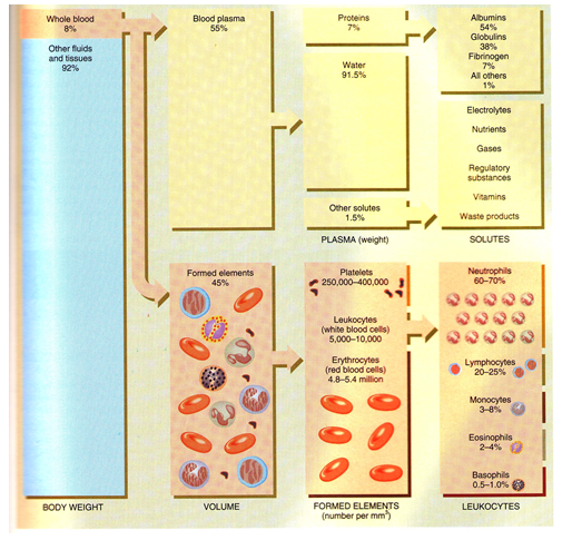
A type of liquid connective tissue that helps in transportation, regulation of pH, body temperature and protection (antibodies). It is composed of two parts. Approximately 55% is blood plasma and 45% is formed elements.
- Study of circulatory system is called as angiology. Father of blood circulation is William Harvey.
- Haematology is study of blood.
- Blood is the softest tissue in body and is derived from mesoderm.
- Blood is slightly alkaline (pH-7.4).
- Blood constitutes 8% of total body weight, blood volume in an average sized male is 5-6 litres and average female is 4-5 litres.
- pH of blood is maintained by balancing the ratio of sodium bicarbonate and carbonic acid. Buffer of blood is sodium bicarbonate.
- Blood is 2.5 times more viscous than water.
- Consists of watery fluid – plasma (55 to 60%), and formed elements – R.B.Cs, W.B.Cs, Platelets (40 to 45%).
- Plasma : Water (91-92%) solids (8-9%) out of which organic solids are 7% (albumin 4.4%, globulin 2.3%, fibrinogen 0.3%) and inorganic solids are 1%.
- Globulins are formed by plasma cells in lymphoid organs; other plasma proteins are formed in the liver.
- Globulins acts as antibodies (Immuno-globulins).
R.B.C. (Erythrocytes)
- Amphibian R.B.Cs are largest amongst vertebrates, those of Amphiuma and Proteus are largest amongst amphibians.
- Mammals have smallest R.B.Cs amongst vertebrates. Musk deer has smallest RBCs amongst mammals and elephant has largest RBCs among mammals.
- Whereas RBCs of other vertebrates are oval and nucleated, those of mammals are roughly biconcave and without nucleus. This increases the surface area of RBCs and enables them to contain more haemoglobin.
- R.B.C count in Human – 5.4 million/mm³ of blood (slightly less in women, more in children).
- In embryonic condition, R.B.Cs are formed in yolk sac and bone marrow (haemopoeitic organ).
- In adult mammal, red bone marrow in long bone is the main site for formation of RBCs.
- RBCs count increases during exercise and stress, and decreases during rest, sleep, menstruation and pregnancy.
- People living in hilly areas have more RBCs. RBCs count sharply falls in anaemia and rises in polycythemia.
Life Span of RBC
- Life span of RBC in Rabbit – 80 days
- In man – 120 days
- In Frog – 100 days
- Radioactive chromium method. It is used for estimation of life span of RBC
ESR
- Erythrocyte sedimentation rate.
- It is measured by Wintrobe's tube. It is rate of settling down of RBC.
Haemocytometer
- It is an instrument used for counting both W.B.Cs. and RBCs.
Structure of RBCs of man
- Biconcave, non-nucleated bounded by Donnan's membrane (plasma membrane of RBC)
- Donnan's membrane is made of Lipoprotein which is 0.2 μ in thickness (mainly lipoid in nature)
- Lipid – 53% Protein – 40% Carbohydrate – 7%
W.B.C. Leukocytes
- Much larger than RBCs but less in number (about 8000/mm³ of blood).
- Ratio RBC : W.B.C. = 700 : 1
Two Main Types:
- Granulocytes (68%): eosinophils, basophils, neutrophils
- Agranulocytes (32%): lymphocytes, monocytes
Granulocytes
- Eosinophils (acidophils) 2.8%, nucleus is bilobed. An abnormal rise in its count is called eosinophilia which generally occurs in infections. Stain dye is eosin. Number also increases in allergy and worm infection, e.g.– Ascaris. These are also known as Acidophils. They play an important role in hypersensitivity. These secrete histamine.
- Basophils least numerous (0.5 to 2%), nucleus is nearly S shaped, functions like mast cells of connective tissue. Both basophils and eosinophils are involved in allergic reaction. Life span = 12-15 days. Nucleus is less distinct. They represent MAST Cells of connective tissue. Number increases in Chicken pox. These Secrete – (i) Histamine – Vasodilator (ii) Heparin – Anti coagulant (iii) Serotonin – Vasoconstrictor.
- Neutrophils most numerous (65%) and most active, nucleus 2 to 5 lobed. They engulf microbes and dead cells. Also known as heterophils. Neutrophils are also known as micropoliceman of blood (10 μ size). Life span = 2-4 days. Nucleus is multilobulated. Phagocytic in nature. Produce hydrolytic enzyme. Number increases in bacterial infection.
Agranulocytes
- Lymphocytes 26%, nucleus large, produce antibodies. Small in size (7 μ). Nucleus is central and rounded. Life span 3 days. Number increases in viral fever and whooping cough. Granules are absent in cytoplasm.
- Monocytes 6%, nucleus-horse shoe shaped, phagocytic, changes into macrophages after entering tissue spaces. Largest WBC (22 μ). It is macropoliceman of blood. Life span = 28 days or one month. Number increases in tuberculosis (T.B.). They represent macrophages of connective tissue.
- Rise in W.B.C. count is called leucocytosis.
- Fall in W.B.C. count is called leucopenia.
- Abnormal rise in W.B.C. count is called leukemia.
- TLC – Total Leucocyte count = Normally 8000-9000 mm³
- DLC – Differential Leucocyte count
- Eosinophils – 2.8% Basophils – 0.2% Neutrophils – 65%
- Lymphocyte – 26% Monocyte – 6%
Blood Platelets
- Thrombocyte like cells.
- Found in mammals only.
- Platelets are derived from megakaryocytes in bone marrow.
- Life span = 10 days.
- Shape is irregular, oval or spherical.
- Platelets are non-nucleated.
- Count 2-5 × 10⁵ cells/mm³ of blood.
- Platelets are the fragments which form as bud and pinch off from certain large cell (mega karyocytes) in bone marrow.
- Blood platelets play an important role in blood clotting.
Spindle Cells or Thrombocytes
- "Spindle cells" occur in all vertebrates other than mammals. Its function is similar to mammalian blood platelets.
- Nucleus is present, oval or spherical in shape.
- Help in blood clotting.
- Clot inside a vessel is called thrombus and its formation is known as thrombosis.
- Thalassemia is an inherited blood disorder resulting in anaemia. The only treatment of thalassemia is bone marrow transplant, has been performed for the first time in India at Christian Medical College Hospital in Vellore.
Life Span of Blood corpuscles
- R.B.C. (120 days in mammal, 100 days in frog), Granulocytes (1 to 8 hrs.) Agranulocytes (few days to a month), platelets – 8 to 10 days.
- 'Spleen' is often referred to as 'graveyard' of RBC (site of RBC disposal)
- W.B.C. mostly get destroyed in connective tissue.