History Of Health And Diseases
- Early Greeks and the Indian Ayurveda believed that health is a balanced state of the body and the mind.
- Health, for a long time, was considered as a state of body and mind where there was a balance of certain ‘humors’
- There was an old reflective thought that persons with ‘blackbile’ belonged to a hot personality and would have fever. William Harvey disproved this good humor-bad humor hypothesis by demonstrating normal body temperature in these people using a thermometer. He also discovered circulation.
- Hippocrates (460-359 B.C.), the great physician, was the first person to write detailed descriptions of diseases symptoms and importance of the need for good diet, fresh air & rest.
- Hippocrates separated medicine from religion, superstition & philosophy and is called ‘The Father of Medicine’.
Concept Of Health
(i) The term “health” means “Wholeness”.
(ii) In 1948, the World Health Organization (WHO) defined ‘health’ as
‘A state of completephysical, mental & social well-being and not merely an absence of disease or infirmity.’ Later, a fourth dimension- spiritual health was added.
(iii) A balanced diet, personal hygiene and regular exercise are important for maintaining good health.
(iv) Today it is also known that mind influences the immune system through the neural and endocrine system. Also, it stated that the immune system maintains one’s health.
(v) Thus, the mind and mental state affect health.
(vi) Since ancient times, yoga has been practiced to achieve physical and mental health.
(vii) Health is affected by
- genetic disorders – deficiencies with which a child is born and deficiencies/defects which the child inherits from parents from birth;
- infections and
- life style including food and water we take, rest and exercise we give to our bodies, habits that we have or lack etc.
Health Organisations
(i) WHO is World Health Organization set up in 1945 with its head office in Geneva.
(ii) It has 6 regional offices in the world, of which the Indian office is in Delhi.
(iii) Red Cross is the oldest health organization in the world and was set up in 1864.
(iv) The other popular global organizations include:
- UNESCO (United Nations Educational, Scientific and Cultural Organization)
- UNICEF (United Nations International Children Emergency Fund)
(v) In every state, the community health services are provided by the regional offices of the Public Health Department (PHD).
CONCEPT OF DISEASE (French for ‘not at ease’)
(i) When the functioning of one or more organs or systems of the body is adversely affected, various signs and symptoms are seen.
(ii) A sign is an indication of a disease observable to the trained eye of a doctor i.e. that which the doctor observes.
(iii) A symptom is an outward manifestation of illness or patients complaints.
(iv) Disease is a disorder in the physical/physiological/psychological functions of body, caused by micro-organisms or nutritional deficiency or genetic disorder or any other reason.
(v) Disease may be acute or chronic.
(vi) An acute disease is a short term illness which tends to start abruptly and may clear up quickly (common cold) or may persist for long time (asthma).
(vii) A chronic disease is always persistent and long-lasting and may be mild or serve.
(viii) Health and disease have no separate existence of their own; they are names given to states or conditions of living things.
CLASSIFICATION OF DISEASES
The study of classification of disease is known as Nosology.
On the basis of occurrence in the body diseases are classified as follows.
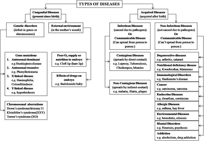
CONGENITAL DISEASES OR HEREDITARY DISEASES:
- (i) These diseases are present at birth.
- (ii) They are passed from parents to child.
- (iii) They are responsible for 50% deaths in new born babies.
- (iv) They can occur due to the following causes:
(1) Single gene mutations:
They can further be classified as
(a) Autosomal dominant diseases:
These are transmitted through autosome and are expressed when only a single copy of mutant gene is present.
E.g. Huntington’s disease, Myotonic dystrophy, Marfan’s syndrome etc.
(b) Autosomal recessive diseases:
Transmitted through autosome and are expressed when both the copies of genes are mutant. e.g. Phenylketonuria, Cystic fibrosis, Alkaptonuria, Albinism etc.
(c) Sex linked diseases:
Transmitted through sex chromosome and are expressed when both the copies of genes are mutant.
E.g. Haemophilia (X-linked disease)
Colour blindness (X-linked disease)
Hypertrichosis (Y-linked disease)
(2) Chromosomal mutations/ aberrations:
These may occur due to structural changes such as deletion, duplication or inversion within a chromosome.
E.g.
- Down’s syndrome (trisomy 21) – has 47 chromosomes
- Patau’s syndrome (trisomy 13) – has 47 chromosomes
- Edward’s syndrome (trisomy 18) – has 47 chromosomes
- Klinefelter’s syndrome (44A + XXY) – has 47 chromosome
- Turner’s syndrome (44A + XO) - has 45 chromosomes
(3) External changes/Environmental effects in embryo:
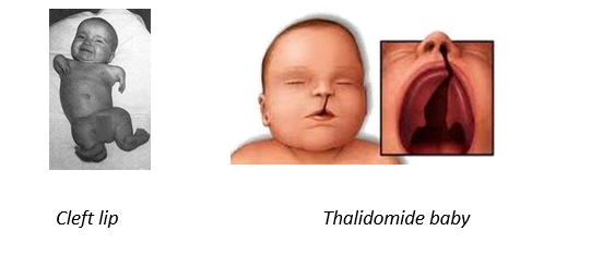
(a) If a pregnant woman takes thalidomide (drug), it severely hampers embryonic growth and baby may have no forearm (thalidomide baby).
(b) Physiological disturbances due to environmental factors during embryonic development may also affect the child. E.g. Cleft lip (hare lip) occurs due to poor O2 supply or poor nutrition to embryo.
ACQUIRED DISEASES:
(i) These diseases develop after birth.
(ii) They are not passed from parents to children.
(iii) They are of two types:
(1) Communicable Diseases (Infectious Diseases):
- The diseases which are transferred from one infected person to other through contact, air, water, food etc. are called communicable diseases.
- They are also called infectious diseases.
- These are pathogenic diseases as these are caused by micro-organism or pathogens(Greek, Pathos = diseases, genes = producing).
- The microorganism may be bacteria, virus, fungi, protozoa etc.
Communicable Diseases are further classified into two types:
(a) Contagious Diseases:
In these diseases, pathogen spreads just by direct contact between infected person and other person (through touch, cloths, sneezing, coughing spitting etc.) E.g. Leprosy, Tuberculosis, Chickenpox, Measles etc.
(b) Non Contagious Diseases:
In these diseases, pathogen does not spread by direct contact but requires a transport agency.
The transport agency may be a
- (i) vector (biotic factor) like mosquito, housefly, bugs etc. or
- (ii) vehicle (abiotic factor) like contaminated food, water and blood
E.g. Hepatitis, malaria, plague, typhoid, cholera, tuberculosis etc.
(2) Non Communicable Diseases (Non-infectious Diseases):
These diseases are confined to the person who suffers and do not get transferred to others by air, water, food or any mean. They are also called non-infectious diseases.
These are the diseases which are caused by any agent other than pathogen hence they are also called non-pathogenic diseases.
Depending upon the causative agent, they are further classified into many types as
(a) Environmental Diseases:
Industries release toxic chemical substances which causes diseases.
E.g. Bronchitis, silicosis, asbestosis, Minamata disease (mercury poisoning),
Itai itai (cadmium poisoning) etc.
(b) Allergic Diseases:
They are caused by hypersensitivity of body to certain external agents (allergens)
E.g. Asthma, hay fever (caused by pollens), anaphylaxis caused by bee sting etc.
(c) Mental Disorders:
They affect the central nervous system (CNS).
E.g. Psychosis, epilepsy, neurosis etc.
(d) Degenerative diseases (ageing):
They are caused by malfunctioning of important organs like lungs, heart, joints etc. due to ageing. E.g. Osteoarthritis, arteriosclerosis, cataract
(e) Nutritional deficiency diseases:
Some of the nutritional deficiency diseases are as follows:
| No. | Deficiency of | Disease |
| 1. | Protein | Kwashiorkor |
| 2. | Protein Energy Malnutrition (PEM) | Marasmus |
| 3. | Iodine | Goiter |
| 4 | Fluorine | Skeletal and Dental fluorosis |
| 5. | Vit A (retinol) | Xeropthalmia, Night blindness,
Xeroderma |
| 6. | Vit B1 (thiamine) | Beri Beri |
| 7. | Vit B2 (riboflavin) | Cheliosis |
| 8. | Vit B3 (Niacin) | Pellagra |
| 9. | Vit B12 (cyanocobalamin) | Pernicious anaemia |
| 10. | Vit B12 and folic acid | Megaloblastic anaemia |
| 10. | Vit C (ascorbic acid) | Scurvy |
| 11. | Vit D (calciferol) | Rickets in children,
Osteomalacia in adults |
| 12. | Vit E (tocopherol) | Sterility |
| 13, | Vit K (phylloquinone) | Bleeding and
Hypothrombinemia (clotting disorders) |
(f) Endocrine diseases:
Some of the endocrine diseases are as follows:
| No. | Cause | Disease |
| 1. | Deficiency of growth hormone in children | Dwarfism |
| 2. | Hypersecretion of growth hormone in children | Gigantism |
| 3. | Deficiency of growth hormone in adults | Simmond’s Disease |
| 4. | Hypersecretion of growth hormone in adults | Acromegaly |
| 5. | Deficiency of thyroxine in children | Cretinism |
| 6. | Deficiency of thyroxine in adults | Myxoedema |
| 7. | Hypersecretion of thyroxine | Grave’s disease |
| 8. | Deficiency of glucocorticoids from adrenal | Addison’s disease |
| 9. | Hpersecretion of glucocorticoids from adrenal | Cushing’s disease |
| 10. | Deficiency of insulin from pancreas | Diabetes mellitus |
| 11. | Deficiency of vasopressin (A.D.H.) | Diabetes insipidus |
| 12. | Deficiency of parathormone | Tetany |
(g) Diseases caused by addictive substances:
Tobacco causes mouth cancer, peptic ulcer and lung diseases like bronchitis, laryngitis, emphysema and chronic cough,
Alcohol causes cirrhosis (damage to liver), gastritis, peptic ulcers, pancreatitis, neuritis, heart disorders and atherosclerosis etc.
Narcotic drugs cause damage to nervous system, respiratory tract and other organs.
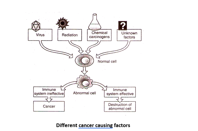
(h) Cancer: It is an uncontrolled, unorganized and abnormal division of cell forming a mass or group of undifferentiated malignant cells..
Some of the cancers are as follows:
| No. | Tissues/ Organs involved | Type of cancer |
| 1. | Epithelial tissue | Carcinoma |
| 2. | Connective and Muscular tissue | Sarcoma |
| 3. | Lymph nodes, Spleen | Lymphoma |
| 4. | Leucocytes of blood | Leukemia |
| 5. | Glands | Adenoma |
(i) Immunological Disorders (Auto-immunity):
A person may develop autoimmunity against tissue, organs or secretions of its own body. E.g. Hashimoto’s thyroiditis: In this disease, antibodies are produced in a person’s body against his own thyroglobulin. In such persons, the glandular tissue of thyroid is lost and is replaced by lymphoid tissue. Rheumatoid arthritis: painful joints
Disease Causing Agents
Any substance which causes disease is called as disease causing agent or etiological factor.
The study of cause of disease is called etiology.
There are 4 major types of disease causing agents as follows:
(1) Physical Agents
(2) Chemical Agents
(3) Mechanical Agents
(4) Biological agents
(1) Physical agents:
Physical agents include exposure to:
- (a) excess heat causing stroke
- (b) excess cold causing frost bite
- (c) radiation causing cancer. E.g. Strontium 90 causes bone cancer.
- (d) excess sound causing impaired hearing.
(2) Chemical agents:
Some chemical compounds are also causative agents of certain diseases. They are of 2 types
- (a) Endogenous Chemical Agents:
Internal chemical agents are produced in the body as a result of derangement of function.
E.g. urea (ureamia), serum bilirubin (jaundice), uric acid (gout), calcium carbonate (kidney stones) etc.
- (b) Exogenous Chemical Agents:
External chemical agents enter the body by inhalation, ingestion or inoculation to cause diseases.
E.g. Environmental pollutants like dust, gases, insecticides, pollen grains, heavy metals.
Addictive substances like tobacco, alcohol and drugs, like etc.
(3) Mechanical Agents:
Exposure to chronic friction and other mechanical forces may result in crushing.
E.g. Sprains, dislocation, fractures etc can be caused by the mechanical forces.
(4) Biological agents (Infectious agents):
(a) Pathogens are micro-organisms like bacteria, viruses, protozoans, helminthes and fungi which cause diseases.
(b) They show properties like infectivity (ability to infect host) and pathogenecity (ability to induce illness).
(c) These pathogens enter the body, produce toxins during incubation period which interferes with the normal functioning of the body and causes diseases.
(i) Bacterial agents:
Unicellular prokaryotic organisms with cell wall are bacteria. Most bacteria make up the normal flora of the host and are harmless. Some like Streptococcus pneumoniae may grow in the throats of healthy people with a harmless commensal relationship.
Only a minority of the bacteria are pathogens that cause infectious diseases. Robert Koch (1976), a German physician showed that anthrax of sheep was caused by bacterial infection. The disease tuberculosis is named Koch’s disease after him.
Robert Koch also established the Koch’s postulates, basically a procedure to determine whether a certain organism is the source of a particular infection. These postulates are:
- Find and isolate pathogen from host
- Grow pathogen in pure culture
- Introducein a new host to produce symptoms
- Isolate pathogen from the new host
Some of the diseases caused by bacteria are:
| No. | Disease | Causative bacteria |
| 1. | Tuberculosis (Koch’s Disease) | Mycobacterium tuberculosis |
| 2. | Leprosy (Hansen’s disease) | Mycobacterium leprae |
| 3. | Tetanus (Lock jaw) | Clostridium tetani |
| 4. | Botulism (Food poisoning) | Clostridium botulinum |
| 5. | Diphtheria | Corynebacterium diphtheria |
| 6. | Pneumonia | Streptococcus pneumonia |
| 7. | Whooping cough (Pertussis) | Bordetella pertussis |
| 8. | Bacillary dysentery | Shigella |
| 9. | Infective diarrhea | Campylobacter jejuni |
| 10. | Typhoid (Enteric fever) | Salmonella typhi, Salmonella paratyphi A and B |
| 11. | Cholera | Vibrio cholera |
| 12. | Syphilis | Treponema pallidum |
| 13. | Gonorrhea (STD) | Neisseria gonorrhoea |
| 14. | Urinary tract infection (UTI)
(women suffer more than men) |
E. coli, Yeast-Candida, Kleibseilla |
| 15. | Anthrax | Bacillus anthracis |
| 16. | Plague | Yersinia anthracis |
| 17. | Boils and pimples (Acne) | Staphylococci |
(ii) Protozoan Agents:
Unicellular eukaryotic micro-organisms that lack cells walls, are called protozoans.
Some of the diseases caused by protozoans are:
| No. |
Disease |
Causative protozoan |
| 1. |
Amoebic dysentery (amoebiasis) |
Entamoeba histolytica |
| 2. |
Pyrrhoea (gum disease) |
Entamoeba gingivalis |
| 3. |
African Sleeping sickness |
Trypanosoma gambiense |
| 4. |
Kala azar/ Leishmaniasis/ Oriental sore |
Leishmania donovani |
| 5. |
Malaria |
Plasmodium vivax |
(iii) Viral agents:
Viruses are obligatory parasitic microorganisms without cells. They have nucleic acid with a protein coat. They are on the threshold of life. They are highly host specific.
Some of the diseases caused by viruses are:
| No. | Disease | Causative virus |
| 1. | Common cold | Rhino virus |
| 2. | Swine flu | H1N1 virus |
| 3. | Influenza | Orthomyxovirus |
| 4. | Mumps (Infectious parotitis) | Paramyxovirus |
| 5. | Yellow fever | Flavi virus |
| 6. | Chickenpox in children/
Shingles in adults |
Varicella-zoster |
| 7. | Small pox (Variola) | Variola virus |
| 8. | Measles (Rubeola) | Morbilli virus |
| 9. | Poliomyelitis | Polio virus |
| 10 | Rabies (Hydrophobia) | Rhabdo virus |
| 11. | AIDS | HIV (Retrovirus) |
| 12. | Hepatitis- A, B, C, D and E | Hepatitis virus- A, B, C, D & E resp. |
| 13. | Burkitt’s lymphoma | Epstein Barr virus |
(iv) Helminthes agents:
Helminthes may be flatworms or round worms which cause diseases.
Some of the diseases caused by helminthes are:
| No. | Disease | Causative helminth |
| 1. | Taeniasis (Neurocysticercosis) | Taenia solium-pork tapeworm
Taenia saginata-beef tapeworm Parasites of human intestines |
| 2. | Ascariasis | Ascaris lumbricoides |
| 3. | Filariasis (Elephantiasis) | Wuchereria bancrofti |
| 4. | Fascioliasis
diarrhea, anaemia, liver damage, lowered body resistance. |
Fasciola hepaticus (Liver fluke)
Its alternative host is a snail. |
| 5. | Schistosomiasis | Schistosoma mansonii |
| 6. | Hook worm infection | Ankylostoma duodenale |
(iv) Fungal agents:
Fungi are eukaryotic, multicellular saprophytic organisms that can cause disease.
Fungi cause disease through 3 mechanisms:
- (a) Immune response: Causing allergic reaction e.g. Aspergillus
- (b) Producing endotoxins (called mycotoxin) e.g. Liver cancer is caused by aflatoxin produced by Aspergillus flavus.
- (c) Through infections causing mycosis.
Some of the diseases caused by fungi are:
| No. | Disease | Causative fungi |
| 1. | Ring worm (Tinea corporis) |
Dermatophytes like species of Trichophyton, species of Epidermophyton and species of Microsporum. |
| 2. | Athelet’s foot (Tinea pedis) | |
| 3. | Genital itch (Tinea cruris) | |
| 4. | Barber’s itch (Tinea barbae) | |
| 5. | Onychomycosis (Tinea ungium) | |
| 6. | Actinomycosis
(abscesses in mouth and lungs) |
Species of Actinomyces |
| 7. | Blastomycosis
(Gilchrist’s disease) |
Species of Blastomyces dermatitidis |
| 8. | Coccidiomycosis
(California fever) |
Species of Coccidioides |
(v) Rickettsial agents:
(i) Rickettsias are prokaryotes included in proteobacteria and are intermediate between viruses and bacteria.
(ii) They are gram negative, oval to rod shaped, non-motile, and obligatory intracellular
parasites.
(iii) Most of them live in the intestines of insects.
(iv) Rickettsias are mainly diseases caused by various rickettsial organisms, as follows:
- (a) Typhus fever (by Rickettsia prowazeki which is louse borne)
- (b) Rocky mountain spotted fever
- (c) Query fever (Q-fever)
- (d) Trench fever
Modes Of Transmission Of Diseases
Communicable diseases may be transmitted from an infectious person to a susceptible healthy person. The mode of transmission of these diseases depends on infectious agent,way of entry and
local ecological conditions.
The study of mode of transmission of disease is called epidemiology.
There are two modes of transmission:
(1) Direct transmission
(2) Indirect transmission
(1) Direct Transmission:
In this type, pathogens are directly transmitted without an intermediate agent.
Direct transmission can take place by droplet infection, direct contact, soil contact animal bite and
through placenta.
(a) Droplet Infection:
The direct exposure of healthy person to the droplets of saliva and nasopharyngeal secretions during coughing, sneezing, speaking and spitting of infected person. These are more common in crowded & congested living conditions.
e.g. Tuberculosis, Diphtheria, Mumps, Common cold, Whooping cough, Pneumonia etc
(b) Direct Contact:
Contagious diseases are directly transmitted from source to susceptible individual by hand shake, mouth to mouth kissing, prolonged close contact and intimate sexual contact.
e.g. Sexually transmitted diseases, leprosy, skin and eye infections, Ringworm,Chicken pox, Small pox, measles etc
Sexually Transmitted Diseases
(i) Sexually transmitted diseases (STD) are also known as venereal diseases.
(ii) These are diseases which are predominantly spread by sexual contact.
Some of the sexually transmitted disease are:
| No. | Disease | Causative organism |
|
Bacterial STD |
||
| 1. | Syphilis | Treponema pallidum (spirochetes) |
| 2. | Gonorrhoea | Neisseria gonorrhoeae |
| 3. | Chancroid | Haemophilus ducreyi |
| 4. | Vaginitis | Chlamydia trachomatis |
|
Viral STD |
||
| 5. | Herpes genitalis | Herpes simplex virus |
| 6. | Condyloma acuminatum | Human papilloma virus |
| 7. | Molluscum contagiosum | Molluscum contagiosum virus |
| 8. | AIDS | HIV-1 and HIV–2 |
|
Protozoal STD |
||
| 9. | Trichomoniasis | Trichomonas vaginalis |
(c) Contact with Soil:
The infection acquired by direct exposure to the disease agent in soil, compost or decaying vegetable matter in which it normally leads a saprophytic life.
e.g. Hookworm larvae, Tetanus bacteria, Mycosis causing fungi etc.
(d) Animal Bite:
Animal bite causes inoculation of diseases causing agent directly into the skin & mucosa.
e.g. Rabies or hydrophobia in man is caused by a virus that is transmitted through a dog bite. The virus is present in the saliva of the rabid animal like dogs, cats, bats, monkeys etc. and enters the healthy person through the wound. It is the only communicable disease of man which is fatal if not treated immediately after infection.
Psittacosis (parrot fever) spreads to human beings through contact with infected birds.
(e) TransplacentalTransmission (Vertical Transmission):
Direct transmission of disease agent through mother’s placenta to the foetus can also cause disease.
e.g. Syphilis, Rubella, Toxoplasma, Hepatitis-B, Cytomegalovirus, Herpes virus, Varicella virus and AIDS
(2) Indirect Transmission:
Pathogens are transmitted from infected to healthy individual through intermediate agents.
This transmission includes five F’s – flies, fingers, formites, food and fluid.
(a) Vector Borne Diseases:
Vectors or carriers are those animals which carry the pathogens from an infected person to another potential host.
Some of the vectors that cause diseases are:
| No. | Vector | Scientific name | Disease in humans |
| 1. | House fly | Musca domestica | Cholera, typhoid etc. |
| 2. | Female Anopheles mosquito | Anopheles | Malaria |
| 3. | Female Culex mosquito | Culex | Filariais (elephantiasis) |
| 4. | Female Aedes mosquito | Aedes | Yellow fever, Dengue |
| 5. | Sand fly | Phlebotomus | Kala Azar and oriental sore |
| 6. | Rat flea | Xenopsylla cheopsis | Bubonic plague |
| 7. | Tse-tese fly | Glossina | Africa sleeping sickness |
| 8. | Ticks | Dermacentor | Rocky mountain spotted fever |
| 9. | Reduviid bug | Rhodnius & Triatoma | Chagas disease in South America |
| 10. | Body louse | Pediculus humanis | Endemic typhus
Epidemic typhus |
| 11. | Itch mite | Sarcoptes scabiei | Scabies |
(b) Vector Borne Diseases:
Transmission of infectious agents through the agency of water, food, blood or semen causes diseases.
e.g. Jaundice, Hepatitis B, AIDS, Polio etc.
Raw cow’s milk may contain bovine tuberculosis bacterium.
Meat of pig infected with Tapeworm is called measly pork.
(c) Air Borne Diseases:
Diseases caused by particles of droplet nuclei and dust in air through coughing, sneezing and spitting of infected person.
(d) Fomite Borne Diseases:
Contamination of articles of daily use can cause transfer of infectious agents between the healthy person and patient suffering from the respective diseases. These articles are referred to as fomites. e.g. handkerchief, towels, clothes, utensils, toys, door handles, taps etc. In a hospital these also include, bed linen, covers, dressing materials, syringes and needles.
Pseudomonas aeruginosa can spread by direct contact through infected bed linen, instruments etc. (It is one of the commonest agents for hospital acquired infections).
Other diseases that can be caused by fomites are Diphtheria, Typhoid, Leprosy, Hepatitis-A etc
(e) Unclean Hands and Fingers:
This is may cause direct transmission of pathogens from hand to mouth.
e.g. Ascariasis, Typhoid, Hepatitis-A etc.
Some Basic Concepts of Diseases
(i) Source of infection: The person, animal, object or substance from which an infectious agent to the host.
(ii) Reservoir of infection: A reservoir is any person, animal, arthropod, plant, soil or substance in which an infectious agent lives and multiplies
(iii) Endemic: Infectious disease always present in a region. E.g. Malaria in India
(iv) Epidemic: Infectious disease affecting a community appearing suddenly. E.g. Conjunctivitis, cholera
(v) Pandemic: Disease affecting several places throughout the world. E.g. AIDS, Diabetes
(vi) Infection: Successful entry and multiplication of the pathogen inside the host body.
(vii) Infestation: Parasitic diseases caused by animals such as arthropods (i.e. mites and ticks),
lice, and worms. E.g. Infestation of the head-louse
(viii) Chain of Infection: Source of infection, Mode of transmission and Suspectible host together form a chain of infection
(ix) Noscomial Infection: Infection acquired by the patient during his stay in the hospital or health care facility.
(x) Iatrogenic disease : Disease caused due to any medical procedure by the physician.
(xi) Incubation period : Time between entrance of the pathogen and appearance of symptoms.
(xii) Parasitology : Study of causative organism and its life cycle.
(xiii) Pathology: Branch of medicine dealing with cause, effects, mechanism, nature
and evolution of disease. It deals with morphology and functional changes in the body.
(xiv) Prophylaxis: Preventive measures of the disease.
(xv) Therapy: Method of treatment of the disease.
STUDY OF VARIOUS DISEASES
A few representative members from different groups of pathogenic organisms are discussed here
| No. | Disease | Causative organism | Group of organism |
| 1. | Typhoid | Salmonella typhi | Bacteria |
| 2. | Pneumonia | Streptococcus pneumoniae & Haemophilus influenzae | Bacteria |
| 3. | Common cold | Rhino virus | Virus |
| 4. | Malaria | Plasmodium vivax | Protozoa |
| 5. | Amoebiasis | Entamoeba histolytica | Protozoa |
| 6. | Ascariasis | Ascaris lumbricoides | Ascehelminthes |
| 7. | Filariasis | Wuchereria bancrofti and Wuchereria malayi | Ascehelminthes |
| 8. | Ringworm | Microsporum, Trichophyton and Epidermophyton | Fungi |
Typhoid (Enteric fever)
Typhoid is an acute, febrile, infectious disease.
Typhoid fever has received various names, such as gastric fever, abdominal typhus, infantile remittent fever, slow fever, nervous fever, pathogenic fever, etc. Typhoid was discovered by Bretonneau in 1826. The name “typhoid” was given by Louis in 1829, as a derivative from typhus.
Causative agent of typhoid:
Typhoid is mainly caused by Salmonella typhimurium or Salmonella typhi.
Other species causing typhoid are Salmonella paratyphi A and Salmonella paratyphi B.
Properties of Salmonella typhimurium:
(i) Salmonella typhi was observed by Eberth in 1880 and isolated by Gaffky in 1884.
(ii) These bacteria belong to family Enterobacteriaceae as they reside in digestive system of host.
(iii) They are rod shaped, facultative anaerobes, 2-4 µ in length and 0.5-0.8 µ in width.
(iv) They are motile and possess multiple flagella (Peritrichous flagella).
(v) It is a Gram negative bacteria.
(vi) Its pathogenicity is due to an outer membrane consisting largely of lipopolysaccharides (LPS) which protect the bacteria from the environment.
(vii) The LPS is made up of
- (a) O-antigen, a polysaccharide core
- (b) Lipid A is made up of two phosphorylated glucosamines which are attached to fatty acids. These phosphate groups determine bacterial toxicity.
(viii) Animals carry an enzyme that specifically removes these phosphate groups in an attempt to protect themselves from these pathogens.
(ix) The O-antigen, being on the outermost part of the LPS complex is responsible for the host immune response and makes it difficult for antibodies to recognize.
(x) These bacteria contain 3 antigens:
- (a) Flagellar ‘H’ antigen
- (b) Somatic ‘O’ antigen on cell wall
- (c) ‘V’ antigen on the capsule polysaccharide (unique to S.typhi).
(xi) O and H antigen produce different antibodies in humans at different rates.
(xii) ‘H’ antigen induces slow antibody formation.
(xiii) S.typhi does not produce endotoxins unless faced with destruction.
(xiv) The bacilli penetrate the mucosa of the ileum after ingestion and pass via the lymphatic system to the mesenteric lymph nodes.
(xv) After the period of multiplication, they pass through the thoracic duct into the bloodstream infecting the liver, gall bladder, spleen, kidney and bone marrow tissue.
(xvi) From the gall bladder these is further invasion of the Payer’s patches in the intestine and other lymphoid tissue.
(xvii) There is an inflammatory reaction followed by necrosis, sloughing and ulcers.
Modes of transmission of typhoid:
(i) It has faeco-oral route of transmission.
(ii) It spreads through unhygienic conditions and overcrowding.
(iii) It spreads through
(i) Direct transmission: by contaminated hands
(ii) Indirect transmission:
(a) Through contaminated food - like green vegetables and shell fishes which come from water contaminated with human excreta.
(b) Through contaminated water and milk.
(c) Vectors like house flies.
(iv) Carriers:
Individuals not showing symptoms of typhoid but carrying the typhoid bacillus within their body for years are called carriers. They can be source of infection.
Carriers are of two types:
(a) Chronic carriers: Bacilli present in faeces for over 1 year after clinical infection.
(b) Temporary carriers (Convalescent carriers): These individuals are in the recovery stage and bacilli are present in faeces for 6-8 weeks. Typhoid disease is common in males while incidence of carrier stage is greater in females.
Typhoid Mary (Carrier)
(i) Mary Mallon, a typhoid carrier for 32 years is a notorious example.
(ii) She was a chef in a popular New York restaurant.
(iii) She caused at least 7 typhoid outbreaks over 15 years, affecting 1300 people.
(iv) As a result she was nicknamed Typhoid Mary.
(v) She was imprisoned for 23 years to prevent occurrence of infections.
Incubation period of Typhoid: The incubation period of Typhoid is about 10-14 days.
Signs and symptoms (Clinical features) of Typhoid:
Slow progressive fever with bradycardia are characteristic symptoms of typhoid.
1st week symptoms:
(i) Anorexia (loss of appetite).
(ii) Nausea, vomiting, abdominal discomfort and diarrhoea accompanied by green stools
(iii) High grade step ladder fever (1040C, higher in the evening), chills and frontal headache.
(iv) Dryness and coating of tongue but with a clean tip.
(v) Backaches, aches of the limbs and generalized weakness.
(vi) Teeth and lips show brownish deposits.
(vii) Bleeding from nose (Epistaxis) is seen in some cases.
2nd week symptoms:
(i) The headache subsides but there is a rise in fever and the patient may suffer from convulsions.
(ii) Spleenomegaly- enlargement of spleen .
(iii) Blood pressure decreases.
(iv) The skin turns dry and rose coloured skin eruptions appear.
(v) Many patients may suffer from bronchitis.
Thus the early stages of typhoid can be misdiagnosed as pneumonia.
3rd week symptoms:
(i) There is increased chance of intestinal hemorrhage and intestinal perforation.
(ii) Occurrence of intestinal hemorrhage is indicated by a sudden drop in temperature, a weak, rapid pulse and dark bowel discharge.
(iii) After the 3rd week, severe cases show pneumonia, myocarditis, meningitis, encephalitis, neuritis, arthritis and hemolytic anaemia.
(iv) If left untreated, patient may pass into a coma and die.
Investigations for Typhoid:
(i) Blood count: There is leukopenia, a decrease in the number of circulating white blood cells, with eosinopenia and relative lymphocytosis.
(ii) Urine culture: Bacteria are present in urine in 3rd week.
(iii) Blood culture: Turbidity indicates positive test.
(iv) Stool culture: Bacteria is present in faeces in the 2nd week.
(v) Widal Test: Test for antibodies devised by Georges Fernad I. Widal in 1896. It is positive only after 15-23rd day of infection.
Prevention of Typhoid:
There are two vaccines currently recommended by the World Health Organization for
the prevention of typhoid :
(i) Live, oral Ty21a vaccine (sold as Vivotif Berna)
(ii) Injectable Typhoid polysaccharide vaccine (sold as Typhim Vi or Typherix).
Treatment of typhoid:
Antibiotics used are Chloromyecetin (chloramphenicol) and ampicillin.
Common cold
The common cold also known as nasopharyngitis, acute viral rhinopharyngitis, acute coryza or Rhinitis acuta catarrhalis) is a viral infectious disease of the upper respiratory system. It is a highly contagious infection which commonly spreads through nasal secretions. Common cold is the most frequent infectious disease in humans with
(i) the average adult contracting 2 - 4 infections in a year and
(ii) the average child contracting between 6–12 infections in a year.
Causative agents of common cold: Rhinoviruses and corona viruses.
Signs and symptoms or clinical features of common cold:
Common symptoms include
(i) cough
(ii) nasal congestion and discharge
(iii) sore throat & hoarseness of voice
(iv) fever
(v) headache
Sometimes this may be accompanied by
(i) conjunctivitis (pink eye)
(ii) muscle aches and fatigue
(iii) shivering
Treatment of Common cold:
There is currently no known treatment that shortens the duration. Symptoms usually resolve spontaneously in 7 to 10 days, with some symptoms possibly lasting for upto three weeks.
Pneumonia
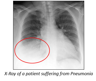
Pneumonia is an inflammatory condition of the lung, especially inflammation of the alveoli or when the lungs fill with fluid (called consolidation and exudation).
Causative agents of Pneumonia:
(i) There are many causes of pneumonia.
(ii) Infection is the most common cause
(a) bacteria like Streptococcus pneumonia and Haemophilus influenza
(b) viruses like influenza virus, respiratory syncytial virus (RSV), adenovirus and Para-influenza.
(c) fungi or (d) parasites.
(iii) Chemical burns or physical injury to the lungs can also produce pneumonia.
Signs and symptoms or clinical features of Pneumonia:
(i) Pneumonia is a common disease that occurs in all age groups.
(ii) It is a leading cause of death among the young, the old, and the chronically ill.
(iii) People with infectious pneumonia often have a cough producing greenish or yellow sputum or phlegm.
(iv) High fever that may be accompanied by shaking chills.
(v) Shortness of breath
(vi) Sharp or stabbing chest pain during deep breaths or coughs. Less frequent symptoms of pneumonia include:
(vii) Coughing up blood
(viii) Headaches
(ix) Sweaty and clammy skin, loss of appetite, fatigue, blueness of the skin, nausea, vomiting, mood swings and joint pains or muscle aches.
Treatment of Pneumonia:
Drugs useful for treatment are erythromycin, tetracycline and sulphonamide.
Amoebiasis (amoebic dysentery):
Amoebiasis is an infection caused by Entamoeba histolytica. Amoebiasis is estimated to cause 70,000 deaths per year worldwide.
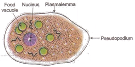
Causative agent of Amoebiasis:
Entamoeba histolytica is a unicellular protozoan.
It is present in the human body in an active form called trophozoite and outside the human body in a protected form called cyst.
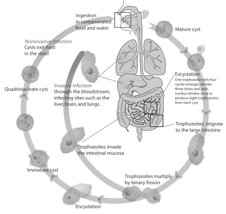
Life cycle of Entamoeba histolytica
Entamoeba histolytica is a monogenetic parasite (single host life cycle, i.e., humans).
Thus humans are definitive hosts for this worm.
(ii) They are transmitted through the faeco-oral route via contaminated food and water.
(iii) Houseflies act as vectors.
(iv) They are ingested in the form of quadric-nucleate cyst.
(v) The cysts reach the small intestine where excystation (breakdown of cyst) takes place to release trophozoites. One cyst produces 8 trophozoites.
(vi) The trophozoites migrate to large intestine where the trophozoites multiply by binary fission.
(vii) These trophozoites may undergo any one of the following two processes:
- (a) Some of these trophozoites invade the intestinal mucosa, reach the blood stream and reach the various organs like liver, brain and lungs causing abscess.
- (b) The remaining trophozoites undergo the process of encystation (formation of cyst) to form a quadric-nucleate cyst which is released in the stools of the patient.
Modes of Transmission of Amoebiasis:
(i) Infection spreads only through the cyst form of the parasite.
(ii) If the parasite is non-encysted, or in the trophozoite form, they usually die in the stomach because of the high acidity. Hence it isn’t infectious.
(iii) The mode of transmission is by faeco-oral route.
(iv) The transmission is indirect by:
- (a) Vehicles like contaminated food or water.
- (b) Transmission by contact with unclean hands and fingers.
- (c) Carrier: An infected individual who himself is not suffering from any symptoms but is capable of transmitting the disease to others through poor hygienic practices.
Incubation period of Amoebiasis:
Can be anywhere between 10 hours to 7 days, usually 2 to 3 days. Majority of cases clear up after 2 to 3 days but some patients may be ill for 2 to 3 weeks. The onset is usually abrupt with fever followed by vomiting, abdominal pain
Signs and Symptoms (Clinical features) of Amoebiasis:
(i) Few individuals suffering from amoebiasis may not be symptomatic and the infection can remain latent in an infected person for several years. They are called carriers.
(ii) There are two types of symptoms; acute (early symptoms) and chronic (prolonged)
(iii) Acute symptoms include:
- (a) Stomach cramps (colic)
- (b) Painful passage of stools (tenesmus)
- (c) Diarrhoea but often blood and mucous in the stool (“amebic dysentery”)
- (d) Nausea
- (e) Weight loss
(iv) Chronic symptoms include:
- (a) Putrid (rancid) stools with blood
- (b) Diarrhoea and constipation may alternate
- (c) Invasive / fulminant amoebiasis causes amoebic dysentery/amebic colitis.
- (d) Recurrent attack of dysentery with gastrointestinal disturbance.
- (e) Abscess in the liver, lung, spleen or brain
- (f) Cutaneous amoebic lesions
- (g) Peritonitis (inflammation of the peritoneum).
Treatment of Amoebiasis:
Amoebiasis can be cured by drugs like Metronidazole or Albendazole.
Ascariasis:
It is caused by parasitic round worm Ascaris lumbricoides belonging to phylum Aschelminthes.
Causative organism of Ascariasis
(i) Ascariasis is caused by Ascaris lumbricoides (round worm of man).
(ii) It belongs to the phylum Aschelminthes (Nemathelminthes).
(iii) It is the most common worm found in human.
(iv) It is a long, thin work tapering at both ends.
(v) It is the largest of the intestinal nematodes parasitizing humans
(vi) They have a cellular or syncytial epidermis.
(vii) The epidermis secretes a thick extra cellular cuticle, covering the body.
(viii) It protects the worm from various secretions of the host.
(ix) The digestive system is complete with 2 openings: terminal anterior mouth end and sub terminal posterior anus.
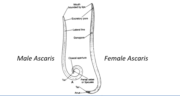
(x) The worm shows sexual dimorphism.
- (a) The female worm is long and straight.
- (b) The male worm is short and curved. It shows the presence of posterior penile setae.
Life cycle of Ascaris lumbricoides:
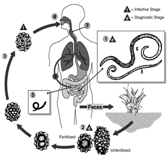
It is monogenetic (involves only one host)
- Adult worms live in the lumen of the small intestine. A female may produce approximately 200,000 eggs per day, which are passed with the faeces.
- Unfertilized eggs may be ingested but are not infective. Fertile eggs embryonate and become infective after 18 days to several weeks i.e., second stage juvenile.
- Fertilized eggs embryonate depending on the environmental conditions (optimum: moist, warm, shaded soil) to become infective.
- Infective eggs are swallowed and larvae hatch.
- Invade the intestinal mucosa, and are carried via the portal, then systemic circulation and/or lymphatics to the lungs.
- The larvae mature further in the lungs(10 to 14 days), penetrate the alveolar walls, ascend the bronchial tree to the throat, and are swallowed.
- Upon reaching the small intestine, they develop into adult worms Between 2 and 3 months are required from ingestion of the infective eggs to oviposition by the adult female. Adult worms can live for 1 to 2 years.
Modes of Transmission of Ascariasis:
(i) Infection spreads only through the fertilized eggs of the parasite.
(ii) If the eggs aren’t fertilized, it is not infectious.
(iii) The mode of transmission is by faeco-oral route.
(iv) Water and vegetables that are contaminated with the infected fertilized eggs are the primary source of infection
(v) The transmission is indirect by:
- (a) Vehicles like contaminated food or water.
- (b) Transmission by contact with unclean hands and fingers.
- (c) Not washing the vegetables and fruits thoroughly before consumption.
Incubation period of Ascariasis:
The symptoms of larval Ascariasis occur 4-16 days after the infection.
Signs and Symptoms (Clinical features) of Ascariasis:
As the adult worms live in the small intestine, the various symptoms include:
(i) Presence of live worms in stools.
(ii) Gastrointestinal pain and discomfort.
(iii) Vomiting, indigestion and diarrhoea.
(iv) A bolus (ball) of worms may also obstruct the intestine leading to colic pains.
(v) Fever
(vi) Internal bleeding and anaemia Due to migration of larvae the patient may show
(vii) Pulmonary symptoms like pneumonitis.
(viii) Neurological symptoms
(ix) Eosinophilia- increased number of eosinophils (WBCs) in blood.
Treatment of Ascariasis
Pharmaceutical drugs like Ascaricides – Mebendazole and Albendazole are used to kill roundworms.
Filariasis
Filariasis is a disease and is a tropical, infectious and parasitic disease caused by thread-like filarial nematodes.
Filariasis is divided into 3 groups (according to the area within the body that they occupy) as follows
- (i) lymphatic filariasis,
- (ii) subcutaneous filariasis.
- (iii) serous cavity filariasis.
Causative agents of Filariasis
Filariasis is caused by thread like filarial nematodes belonging to the Phylum Aschelminthes in the superfamily Filarioidea, also known as “filariae”. There are 9 known filarial nematodes which use humans as their definitive host. Lymphatic filariasis is caused by the worms Wuchereria bancrofti, Brugia malayi and Brugia timori.
Life cycle of filarial worms
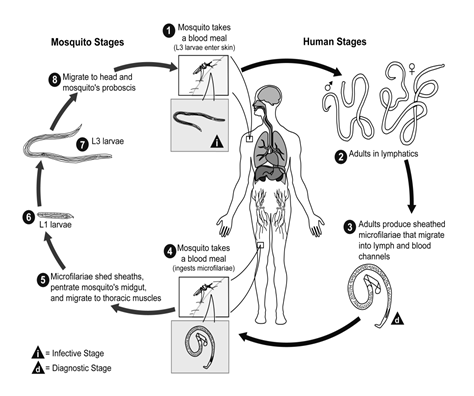
- Human filarial nematode worms have a complicated life cycle, which primarily consists of five stages.
- Filarial worms have a digenetic life cycle having two hosts.
- The adult male and female worms commonly inhabit the lymphatic system of human.
- After the male and female worms mate, the female gives birth to thousands of live micro-filariae.
- The micro-filariae are picked up by vector insects like female Anopheles and female Culex mosquitoes during a blood meal.
- In the intermediate host, the micro-filariae moult and develop into 3rd stage (infective) larvae.
- Upon taking another blood meal, the vector insect injects the infectious larvae into the dermis layer of the human skin.
- After about one year, the larvae moult through 2 more stages, maturing into the adult worms.
Modes of transmission of Filariasis
Filariasis spreads to an infected human through vector insects like female Anopheles and female Culex mosquitoes.
For the Filarial worm
- (i) Humans are their primary hosts
- (ii) The vector insects are their intermediate hosts
Incubation period of Filariasis
The symptoms of Filariasis generally occur 8-16 months after the infection.
Signs and Symptoms (Clinical features) of Filariasis:
- (i) The most distinctive symptom of lymphatic filariasis is elephantiasis.
It is oedema (swelling) and thickening of skin and underlying tissues especially in the legs and genitals. It results when the parasites lodge in the lymphatic system and cause a slowly developingchronic inflammation of the organs in which they live for many years.
The excessive swelling of lower limbs giving an elephant leg like appearance hence it is called elephantiasis.
- (ii) Pain & inflammation of lymph nodes, often accompanied with fever.
- (iii) Nausea & vomiting
- (iv) Vulva and breast enlargement (in females)
- (v) Hydrocoele (swelling of scrotum) in males
Treatment of Filariasis
Diethyl-carbamacine 100 mg twice a day for 3 weeks followed by 100 mg twice a day for 5 days every six months.
Malaria
Malaria is a mosquito-borne infectious disease of humans caused by protist Plasmodium. The disease results from the multiplication of Plasmodium within red blood cells. It is widespread in tropical and subtropical regions, including much of Sub-Saharan Africa, Asia and the America.
Causative agent of Malaria
Malaria is caused by Plasmodium also known as Malarial parasite.
The 4 species of Plasmodium that cause malaria are
(1) Plasmodium vivax causes benign tertian malaria
(2) Plasmodium ovale causes mild tertian malaria
(3) Plasmodium falciparum causes malignant tertian malaria (brain fever)-often fatal.
(4) Plasmodium malariae: causes quartan malaria
Life cycle of Malarial parasite
The life cycle of the malaria parasite (Plasmodium) is complicated.
Plasmodium is a digenetic parasite having 2 hosts; Humans & female Anopheles mosquito
- Life cycle of malarial parasite in Anopheles mosquito:
(i) Mosquitoes first ingest the malaria parasite while feeding on an infected human.
(ii) Once ingested, the gametocytes differentiate into male or female gametes and fuse in the mosquito’s gut.
(iii) This produces an ookinete that penetrates the gut lining and produces an oocyst in the gut wall.
(iv) When the oocyst ruptures, it releases sporozoites that migrate to the salivary glands.
(v) The sporozoites are injected into the skin, when the mosquito bites a normal person.
(vi) Only female mosquitoes feed on blood while male mosquitoes feed on plant sap, thus males do not transmit the disease.
- Life cycle of malarial parasite in Human body:
Malaria develops via two phases: an exoerythrocytic and an erythrocytic phase.
- The exoerythrocytic phase involves infection of the liver.
- The erythrocytic phase involves infection of the erythrocytes, or red blood cells.
(i) Exo-erythrocytic phase:
- When mosquito bites a person, sporozoites (infective stage) enter the bloodstream, and migrate to the liver.
- They infect liver cells (hepatocytes), where they multiply into merozoites.
- They rupture the liver cells, and escape into the bloodstream.
(ii) Erythrocytic phase:
Then, merozoites infect red blood cells, where they develop into ring forms, trophozoites and then schizonts which in turn produce further merozoites. Within the erythrocyte, the parasites multiply asexually.
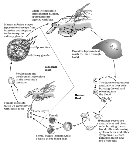
They come out of the erythrocytes by rupturing them and invade fresh red blood cells. The rupture of RBCs is associated with release of a toxic substance, haemozoin, which is responsible for the chill and high fever recurring every three to four days. Several such amplification cycles occur. Thus, classical descriptions of waves of fever arise from simultaneous waves of merozoites escaping and infecting red blood cells.
Mode of transmission of Malaria:
The disease is transmitted to human when an infected Anopheles mosquito bites a person and injects the malaria parasites (sporozoites) into the blood.
Ronald Ross in 1897 explained the relationship between Malaria & Mosquito for which he got Nobel Prize in 1902.
Incubation period of Malaria:
Incubation periods for the various malarial parasites are
(i) 7–14 days for P. falciparum
(ii) 8–14 days for P. vivax and P. ovale and
(iii) 7–30 days for P. malariae
Signs and symptoms (Clinical features) of Malaria:
(i) The classic symptom of malaria is cyclical occurrence of
- Cold stage: It is characterized by a feeling of cold, chills and rigor
- Hot stage: It is characterized by fever
- Sweating stage: It is characterized by profuse sweating which last for 4-6 hours.
This cyclic stages occurs after
- (a) every two days in P. falciparum, P. vivax and P. ovale infections (hence tertian fever)
- (b) every three days for P. malariae (hence quartan fever).
(ii) There may be severe headache and vomiting during fever.
(iii) The patient suffers from hypoglycemia (decreased blood sugar level)
(iv) As erythrocytes are broken down (Haemolysis) in malaria, the patient shows
- (a) Haemoglobinuria- increased excretion of haemoglobin in urine
- (b) Anaemia
- (c) Splenomegaly (enlarged spleen) due to increased haemolysis
- (d) Hepatomegaly (enlarged liver)
(v) The patient may also suffer from arthralgia (joint pain).
(vi) Cerebral malaria caused due to P. falciparum infection may affect the brain causing Cerebral ischaemia, retinal damage and convulsions.
(vii) In severe cases there may be renal failure, coma and even death.
(viii) Blackwater fever is a severe complication of malaria caused due to P. falciparum.
In this the patient releases black urine due to renal failure.
Diagnosis of Malaria:
It is done by preparing a Peripheral smear of blood for detecting the presence of malaria parasite within the erythrocytes
Treatment of Malaria:
(i) Anti-malarial drugs like Quinine (produced from bark of Cinchona officinalis), chloroquine, Primaquin etc.
(ii) Prevention from mosquito bite by using nets, repellants etc.
(iii) Destruction of larva by using Gambusia fish.
Ring worm (Tinea)
Ringworm or Dermatophytosis is a clinical condition caused by fungal infection of the skin in humans, pets such as cats and domesticated animals such as sheep and cattle.
The term “ringworm” is a misnomer, since the condition is caused by fungi of several different species and not by parasitic worms.
Causative agent of Ringworm
- (i) Dermatophytes of the genera Trichophyton, Epidermophyton and Microsporum are the most common causative agents.
- (ii) These fungi attack various parts of the body.
- (iii) The fungi that cause parasitic infection (dermatophytes) feed on keratin, the material found in the outer layer of skin, hair, and nails.
- (iv) These fungi thrive on skin that is warm and moist, but may also survive directly on the outsides of hair shafts or in their interiors.
- (v) In pets, the fungus responsible for the disease survives in skin and on the outer surface of hairs.
Modes of transmission of Ringworm
Infection is generally acquired from soil or by using towel, clothes or even the combs of infected persons. They usually attack skin, hair & nails.
Signs and symptoms or clinical features of Ringworm
(i) Appearance of typical enlarging raised red rings of ringworm which are dry, scaly lesions on various parts of the body such as skin, nails & scalp.
(ii) Lesions are accompanied by intense itching.
(iii) It is named differently according to its position of infection as follows:
| No | Common name | Medical name | Position of lesion |
| (a) | Athlete’s foot | Tinea pedis | feet |
| (b) | Genital itch / jock itch | Tinea cruris | groin |
| (c) | Barber’s itch | Tinea barbae | face |
| (d) | Onychomycosis | Tinea ungium | nails |
| (e) | Ring worm | Tinea corporis | Anywhere else in the body |
(iv) In onychomycosis, nails may thicken, discolour and finally crumble and fall off.
Treatment of ringworm:
Both oral and topical treatment with drugs like terbinafine, fluconazole and itraconazole.
IMMUNE SYSTEM
PRINCIPLES OF IMMUNITY
(i) Immunity is defined as the general ability of the body to recognize, neutralize, destroy and eliminate disease producing agents or resist a particular infection or disease.
(ii) The term immunity was coined by Burnet (Latin immunis: exempt or freedom)
(iii) Immunology is the science which deals with the study of immune system, immune responses to foreign substances and their role in resisting infection.
(iv) The study of structure and function of immune system is called basic immunology.
(v) Other branches of immunology are
- (a) Clinical immunology which includes vaccination/immunization, organ transplantation /organ grafting, blood banking, immunopathology etc.
- (b) Laboratory immunology which includes testing of cellular & humoral immune function.
- (c) Serology which includes Antigen- Antibody reactions.
(vi) Father of immunology is Emil Von Behring who discovered antibodies..
LINES OF DEFENSE
There are three lines of defense mechanisms in the body:
(i) First line of defense:
It is an external defense and prevents entry of micro-organisms in blood.
It contains two barriers:
- (a) Physical barriers: [Structural/anatomical barrier]
It is made up of skin and mucous membrane.
- (b) Chemical barriers: [Functional/physiological barriers]
It includes the secretion from skin and mucous membrane .e.g.
(1) Sweat and sebum maintain acidic pH of skin.
(2) Tears (lacrimal fluid), saliva and mucous secretions from the nose. They contain enzyme called lysozymes which breaks down bacterial cell wall.
(3) Vaginal fluid and gastric fluid which are acidic, killing all the microorganisms.
(ii) Second line of defense:
It is an internal defense and prevents spread of micro-organisms in the body.
(a) Fever (pyrexia): Certain WBCs release pyrogens which increase the temperature of body killing micro-organisms.
(b) Phagocytosis: It is the process of engulfing micro-organisms, directly by cells.
Cells performing phagocytosis are called phagocytes.
There are three phagocytes in the body:
- (1) Neutrophils (granulocytes, WBC).
- (2) Monocytes (agranulocytes, WBC).
- (3) Macrophages (cells of areolar connective tissue).
(c) Anti-microbial substances: The body produces certain chemicals called anti-microbial substances which kill the micro-organisms. E.g. Complements, interferons.
(1) Complements: They are a group of 30 plasma proteins which are generally inactive. They are activated by entry of micro-organisms.
(2) Interferons: They are produced by virus infected cells. Interferons contain antiviral activity and protect neighboring cells from the virus. I and II line of defense are non-specific and are inborn. The above are included in the physiological barriers.
(iii) Third line of defense:
- (a) It is highly specific defense which is not present since birth (acquired).
- (b) It deals with the production of antibodies by lymphocytes.
- (c) Antibodies are highly specific and they will be different for different micro- organisms.
- (d) This line of defense is also called immune system.
- (e) Immune system is made up of blood vascular system & lymphatic system.
Types of IMMUNITY
(i) Immunity is defined as general ability of body to recognize, neutralize, destroy & eliminate disease producing agents or resist a particular infection or disease.
(ii) Immunity is of two types: Innate and Acquired
| - | Innate Immunity | - | Acquired Immunity |
| (i) | It is present since birth | (i) | It is acquired during life |
| (ii) | It is genetically determined | (ii) | It is non genetic |
| (iii) | It is non-specific | (iii) | It is highly specific |
| (iv) | Previous exposure to foreign body is not required | (iv) | Previous exposure to foreign body is required |
| (v) | It doesn’t change with the foreign body | (v) | It changes with the foreign body. Hence it is also called adaptive immunity |
| (vi) | It is the first line of defense | (vi) | It is the last line of defense |
| (vii) | It doesn’t show immunological memory (amnesiatic memory) | (vii) | It shows immunological memory (anamnesiatic immunity) |
| (viii) | It is made up of anatomical barriers, physiological barriers, phagocytotic barriers and inflammatory barriers | (viii) | It is made up of B lymphocytes which provides antibodies & T lymphocytes which provide cellular immunity |
| (ix) | It is seen in all animals | (ix) | It is seen only in vertebrates |
INNATE IMMUNITY
(i) Innate immunity is the inborn capacity of the body to resist pathogens.
(ii) Innate immunity is inborn (present since birth) and is genetically determined.
(iii) It is also called natural immunity.
(iv) It does not depend on previous exposure to foreign substances.
(v) It is non-specific immunity because it is the same for all antigens.
Mechanism of Innate Immunity:
Innate Immunity is brought about by:
- Anatomical barriers
- Physiological barriers
- Phagocytic barriers
- Inflammatory barriers
Natural killer cells (NK cells)
(i) Anatomical Barriers:
Anatomical barriers prevent entry of micro-organisms into the body.
- (a) Skin is made of stratified epithelium which has an outer layer of keratinized-cells called stratum corneum. These dead cells prevent the growth of bacteria.
- (b) Mucous membranes have unicellular goblet glands which produce mucus The mucous membrane also has cilia at many places which drives mucus outside the system. It is present in the epithelial lining of respiratory, gastrointestinal and urinogenital tracts trapping microbes entering the body.
(ii) Physiological barriers:
Physiological barriers prevent the growth of micro-organisms in the body.
These are of 3 types:
Chemical barriers, Fever and Anti-microbial substances
(a) Chemical barriers: They include the secretions from skin and mucous membrane. e.g.
- Sweat and sebum from skin maintain acidic pH of skin. Sebum contains lactic acid and fatty acids which maintains pH of skin between 3 and 5.
- Tears (lacrimal fluid), saliva and mucous secretions from the nose contain hydrolytic enzymes called lysozymes which cause breakdown of bacterial cell wall. Lysozymes are absent in CSF, sweat and urine.
- Vaginal fluid and gastric fluid which are acidic kill all the microbes
(b) Fever (pyrexia):
- Body temperature increases in response to microbes.
- WBC (phagocytes) release substances like interleukin which act as pyrogens (fever producing)
- These interleukins are detected by the hypothalamus which releases prostaglandins.
- Prostaglandins stimulate temperature centre of hypothalamus to increase body temperature.
(c) Anti-microbial substances:
The body produces certain chemicals called anti-microbial substances which kill micro-organisms.
(1) Complements:
They are a group of 30 plasma proteins which are generally inactive.
They are activated by entry of micro-organisms.
(2) Cytokine barrier like Interferons:
They were discovered by Isaacs and Lindemann in 1955. Lymphocytes, macrophages and fibroblasts infected by virus (virus infected cells) produce glycoproteins called interferons. Interferons have anti-viral activity &protect other cells from damage. Interferons have quick but temporary action. They have been effective against influenza and hepatitis.
(3) Transferrins:
Iron binding proteins called transferrins inhibit growth of bacteria.
(4)Basic polypeptides:
Tissues and blood cells form a number of basic proteins that have anti-bacterial property. e.g. Spermine and spermidine which can kill tuberculosis bacteria
(5)Acute phase proteins:
They are plasma proteins that attach to surface of a micro-organism, thus making it more susceptible to phagocytosis by macrophages. Thus they act like opsonins. E.g. C-reactive protein (CRP) and Mannose Binding Protein (MBP)
(iii) Phagocytic barriers:
The phenomenon of phagocytosis was discovered by Metchnikoff.
Phagocytosis is the process of engulfing micro-organisms, directly by cells. Cells performing phagocytosis are called phagocytes.
There are three phagocytes:
(1) Neutrophils: Granulocytes (WBC)
(2) Monocyte: Agranulocytes (WBC)
(3) Macrophage: Cells of areolar connective tissue
Macrophages are also called wandering phagocytes. Other examples of macrophages are Kupffer cells in liver and Clara cells in lungs. In addition to this, monocytes liberated at the site of infection are also converted into macrophages.
(iv) Inflammatory barrier:
- (a) Inflammation is the localized response of the body (organs) to injury (tissue damage) or infection.
- (b) It consists of 5 responses as follows:
(1) Rubor: Redness occurs due to vasodilatation
(2) Tumor: Swelling occurs due to increase permeability of blood vessels
(3) Dolor: Pain occurs due to stimulation of pain receptors
(4) Calor: Heat occurs due to liberation of energy due to breakdown of micro organisms
(5) Loss of function: Inflammation may also cause loss of function of a body part.
- (c) Inflammation is caused due to release of histamine and prostaglandins, by damaged mast cells of connective tissue and basophiles of blood.
- (d) These chemicals dilate and make the blood capillaries more permeable in the region of tissue injury. The vascular fluid comes out of the blood vessels. This fluid contains serum proteins which kill bacteria.
- (e) Inflammation is denoted by adding the suffix ‘– itis’ to the organs e.g. Conjunctivitis (inflammation of the conjunctiva of eye)
(v) Natural killer cells:
(a) About 5-10% lymphocytes in blood are natural killer cells.
(b) They are also present in spleen, lymph node and bone marrow.
(c) They produce hole in the cell membrane of virus infected cells or some tumor cells of body.
(d) Normally lymphocytes are agranular but Natural Killer cells are granular.
(e) Hence they are also called large granular lymphocytes.
Types of Innate Immunity:
There are three types of Innate Immunity as follows:
(i)Species Immunity:
Immunity present in an organism, because it belongs to a certain species.
Species immunity is due to physiological and biochemical differences between tissues of different species.eg.
- (a) Diseases affecting plants and animals generally do not affect human beings i.e.humans are immune to plant diseases.
- (b) Tuberculosis is common in man, cattle, pigs and fowls but it is rare in rabbits, rats and mice
(ii) Racial Immunity:
Racial immunity present in an organism because it belongs to a particular race.
- (a) White human race is more susceptible to skin cancer.
- (b) Black human race is more susceptible to tuberculosis.
- (c) Algerian sheep is more resistant to anthrax than European sheep.
- (d) In Jews, there is racial immunity to TB, Negroes are susceptible to TB.
(iii) Individual Immunity:
Individual immunity is present in a person, due to his/her individual genetic constitution.
e.g. During epidemics of influenza, some people never develop influenza. They have individual immunity.
Acquired IMMUNITY
(i) The resistance or immunity that an individual acquires during life is called acquired immunity (adaptive immunity or specific immunity).
(ii) It is non-genetic and forms the last line of defense.
(iii) Acquired immunity is seen only in vertebrates.
(iv) Acquired immunity forms the immunity system.
(v) The main cells of acquired immunity are lymphocytes which produce antibodies. Unique features of acquired immunity:
(i) Specificity:
It is the ability to differentiate various foreign molecules. Acquired immunity is highly specific. A single antibody is produced against a single antigen.
(ii) Diversity:
Acquired immunity can recognize a vast variety of pathogens or foreign molecules. Hence different antibodies are produced against different antigens.
Thus, antibodies change with change in antigens. Hence, this immunity is also called ‘adaptive immunity’. Approximately, 1020 different types of antibodies are produced by the human body.
(iii) Discrimination between self and non-self:
Acquired immunity can distinguish between body’s own cells (self) and foreign cells and molecules (non-self).
(iv) Memory:
Whenever an antigen enters the body for the first time (first encounter), antibodies are produced to eliminate the antigen.
The memory of this first encounter is stored in the body in the form of chemicals within memory cells.
Hence, during subsequent encounters, antibodies will be formed faster and in greater amount than the first encounter (quicker & stronger immune response).
Types of Acquired Immunity:
Acquired immunity is of two types:
| - |
Passive Immunity |
- |
Active Immunity |
|
(i) |
Immune system is inactive. |
(i) |
Immune system is active. |
|
(ii) |
Antibodies are not produced hence external antibodies are given. |
(ii) |
Antibodies are produced by the immune system against invading pathogens or vaccine. |
|
(iii) |
Immunological memory is not formed. |
(iii) |
Immunological memory is formed. |
|
(iv) |
It is not long lasting. It lasts maximum for 3 months (average 10-15 days). |
(iv) |
It is long lasting for many years. |
|
(v) |
It diminishes (decreases) with the repetition of dose. |
(v) |
It increases with repetition of dose. |
They are further sub classified as:
- Naturally acquired active immunity
- Artificially acquired active immunity
- Naturally acquired passive immunity
- Artificially acquired passive immunity
(1) Naturally acquired active immunity:
It is the Immunity produced when active micro-organisms (pathogens) from nature enter the body i.e. Natural infection. Immune system is stimulated, hence body produces antibodies.
Memory-cells are formed, hence it is lifelong immunity. e.g. a person recovered from measles or chickenpox becomes immune to it for a life time.
(2) Artificially acquired active immunity:
It is the Immunity produced when micro-organisms inactivated by heat and radiation enter the body (vaccination). This artificial preparation of inactive micro-organisms is called vaccine. Vaccines are made up of:
- (a) Dead or alive but attenuated (artificially weakened) pathogens or
- (b) toxoids consisting of microbial components or
- (c) toxins secreted by the pathogens.
Immune system is working, hence body produces antibodies. Memory-cells are formed; hence immunity persists for a long time.
e.g. B.C.G. vaccine against tuberculosis, polio vaccine
Vaccines can be classified as
1st Generation vaccine: Live and weakened or killed forms (Live attenuated vaccines) (Generated both Killer T as well as Helper T responses)
2nd Generation vaccine: Toxoids or recombinant proteins (only Helper T responses)
3rd Generation vaccine: Genetically engineered DNA vaccine containing plasmid (safest)
(3) Naturally acquired passive immunity:
Newborn infants don’t have a well developed immune system.
Their immune system is not working; hence antibodies are not produced by their body.
They are protected by maternal (mother’s) antibodies.
Maternal antibodies reach the child in two ways:
- Before delivery: maternal antibodies (IgG) reach the placenta and enter child’s circulation through the umbilical cord.
- After delivery: maternal antibodies (IgA) reach the child through colostrum/ first breast milk. Memory cells are not produced; hence this immunity is short lived. It lasts for a year.
(4) Artificially acquired passive immunity:
A person suffers from severe disease when his immune system isn’t producing antibodies for that particular disease. Hence external antibodies are given in the form of hyper immune serum or anti-serum. Anti-serum contains antibodies against a particular disease. They may be obtained from humans or other animals e.g. The following antisera are produced from horse (equine)
(i) Anti rabies serum
(ii) Anti tetanus serum
(iii) Anti diphtheria serum
(iv) Anti gas- gangrene serum
(v) Anti venom against snake bite
CELLS OF IMMUNE SYSTEM
The chief cells of acquired immune system are lymphocytes.
A healthy man contains a trillion lymphocytes and about 100 million trillion (1020) antibodies.
Lymphocytes are of two types:
(1) B–lymphocytes:
- ‘B’ in B – lymphocyte stands for Bursa of Fabricius.
- Bursa of Fabricius is lymphoid organ found in birds, located near their cloacal opening.
- Humans do not have bursa.
- Bursa equivalents in humans are bone marrow and foetal liver.
(2) T–lymphocytes:
- (a) ‘T’ in T – lymphocyte stands for thymus.
- (b) Thymus is a bilobed organ located near the heart and beneath the breastbone.
- (c) It size is of a walnut.
- (d) The thymus is quite large at birth but keeps reducing in sizewith age and by the time puberty is attained it reduces to a very small size.
- (e) Thymus is also called ‘Throne of immunity’ or ‘Training school of lymphocytes’.
- (f) The function of thymus is to produce mature T cells.
- (g) Immature thymocytes/prothymocytes leave the bone marrow and migrate to the thymus.
- (h) In thymic education, T-cells that are beneficial to the immune system are spread.
- (i) Those T-cells that might evoke a harmful auto-immune response get eliminated.
- (j) The mature T cells are then released into the blood stream.
The various differentiating points between B and T lymphocytes are:
| - |
B – Lymphocytes |
T – Lymphocytes |
|
(i) |
Formation : They are formed in the red bone marrow from hematopoietic stem cells by a process of haematopoeisis. |
Formation : They too are formed in the red bone marrow from hematopoietic stem cells by a process of haematopoeisis. |
|
(ii) |
Maturation : They mature in the bone marrow, tonsils and Payer’s patches of the intestine. |
Maturation : They migrate to and mature in the thymus with the help of hormone thymosine. |
|
(iii) |
Storage: Mature B cells reside in cortical areas of lymph node. |
Storage: Mature T cells reside in para-cortical areas of lymph node. |
|
(iv) |
B cells form 20–25% of total lymphocytes. |
T cells form 75% of total lymphocytes. |
|
(v) |
B cells have a life span of 5 – 6 days. |
T cells have a life span of 4 – 5 years. |
|
(vi) |
B lymphocytes provide humoral immunity (antibody mediated immunity) It is effective against (i) Extracellular pathogens in (ii) Other antigens in body fluids. |
T lymphocytes provide cell mediated immunity. It is effective against (i) Intracellular pathogens. (ii) Cancer cells. (iii) Foreign tissue transplant |
|
(vii) |
There are two types of B – lymphocytes : (i) Effector B cells /plasma cells: They produce 20 trillion antibodies/day They produce more than 2000 mol of antibodies/sec. They have a characteristic cart wheel nucleus. (ii) Memory B cells: They store information about microbes. They are stored in spleen and lymph nodes. |
There are four types of T – lymphocytes : (i) Helper T cells/T4 cells/CD4 cells : They activate B-cells and killer T cells. (ii) Killer T cells/T8 cells/CD8 cells:: They release cytolysin which forms large holes in target cells. Hence cytolysin is also called perforin. (iii) Suppressor T cells: They stop activity of killer T cells and suppress immune system. (iv) Memory T cells : They store information about microbes. |
CELL MEDIATED IMMUNITY
(i) Cell mediated immunity is provided by T lymphocytes.
(ii) On coming in contact with an antigen, a T-lymphocyte divides rapidly to form a clone of T-cells which are similar in structure but perform different functions.
There are four types of T-lymphocytes:
(1) Helper T cells/ CD4 cells/ T4 cells:
Helper T cells are most abundant T cells. They identify foreign bodies & get sensitized.
Sensitized helper T-cells produce lymphokines to
- (a) Stimulate killer T cells and proliferate other T cells
- (b) Stimulate B lymphocytes
- (c) Attract macrophages to site of infection
(2) Killer T cells/ cytotoxic T cells/ CD8 cells/ T8 cells:
- Killer T cells directly attack and destroy invading microbes, infected body cells and cancer cells.
- Killer T-cell binds to infected cell and secretes perforins/ cytolysin.
- The perforins produce a hole in infected cell.
- It also releases cell killing substances, hence the name cytotoxic T cell.
(3) Suppressor T cells:
These cells suppress entire immune system & prevent it from attacking own body cells.
(4) Memory T cells:
These cells were previously sensitized and retain the sensitization for future. They store information about microbes.
HUMORAL IMMUNITY/ ANTIBODY MEDIATED IMMUNITY
(i) Humoral or antibody mediated immunity is provided by B lymphocytes.
(ii) B-lymphocytes are sensitized directly by antigens as well as by helper T cells.
(iii) On sensitization, a B-lymphocyte divides rapidly to form a clone of B-cells which are similar in structure but perform different functions.
There are 2 types of B-lymphocytes:
(1) Plasma cells/ effector B cells:
The plasma cells produce specialized glycoproteins called antibodies which are passed through body fluids (humor) like blood and lymph. Each antibody is specific for a particular antigen. Antibody molecules may bind to a cell membrane or they may remain free. The free antibodies have three main functions:
- (a) Agglutination of particulate matter, including bacteria and viruses.
- (b) Opsonisation or coating of bacteria to facilitate their subsequent phagocytosis by macrophages
- (c) Neutralization of toxins released by bacteria e.g., tetanus toxin.
(2) Memory B cells:
These cells were previously sensitized and retain their sensitization for future. They store information about microbes.
Lymphoid organs:
The organs in which origin and/or maturation and proliferation of lymphocytes occur are called lymphoid organs. They are of two types: Primary lymphoid organs & Secondary lymphoid organs
(1) Primary Lymphoid Organs:
They are organs where immature lymphocytes differentiate into antigen-sensitive mature lymphocytes.
There are two Primary lymphoid organs- Bone marrow and Thymus The bone marrow is the main lymphoid organ where all blood cells including lymphocytes are produced.
Both bone-marrow and thymus provide micro-environments for the development and maturation of T-lymphocytes.
(2) Secondary Lymphoid Organs:
They are organs where mature lymphocytes migrate to and are stored.
The secondary lymphoid organs provide sites for interaction of lymphocytes with the antigen which then proliferate to form effector cells. The various secondary lymphoid organs are spleen, lymph nodes, tonsils, Peyer’s patches of small intestine and appendix.
Lymphoid tissue located within the lining of the major tracts (respiratory, digestive and urogenital tracts) is called mucosal associated lymphoid tissue(MALT). It constitutes about 50 per cent of the lymphoid tissue in human body. Spleen as a secondary lymphoid organ: The spleen is a large bean shaped organ present on the left side within abdominal cavity.
On a cut section, spleen shows 2 regions:
- (a) Outer red pulp which contains cells called cords of Billroth
The red pulp stores monocytes and macrophages. Thus it acts as a filter of the blood by trapping blood-borne micro-organisms. It is also a large reservoir of erythrocytes and hence called Blood Bank of body.
- (b) Inner white pulp which stores B and T lymphocytes.
Lymph node is a secondary lymphoid organ:
- Lymph nodes are tiny, circular, solid organs scattered along the path of lymphatic vessels.
- Lymph nodes serve to trap the micro-organisms or other antigens, which happen to get into the lymph and tissue fluid.
- Antigens trapped in the lymph nodes are responsible for the activation of lymphocytes present there and cause the immune response.
ANTIGEN
Antigen is defined as any foreign substance invading the body and capable of stimulating an immune response. Antigens are generally large protein molecules. Antigens have following properties:
(1) It has a minimum molecular weight of 5000 Daltons.
(2) Immunogenicity: capacity of antigen to induce an immune reaction
(3) Reactivity: capacity of antigen to react with an antibody. Antigens with both immunogenicity and reactivity are considered complete antigens.
(4) Antigens have a specific region on their surface which acts as an antigenic determinant. It is called epitope. Antibodies identify antigens by epitope.
Incomplete antigens or Haptens
(i) Haptens are non proteinaceous molecules that act as antigens.
(ii) Haptens could be polysaccharides (carbohydrates) or nucleic acids.
(iii) They are never lipids. (Lipids are non-antigenic)
(iv) Haptens themselves can’t stimulate the immune system to produce antibodies but they can chemically combine with a protein to stimulate immune system. Hence haptens are also called incomplete antigens.
ANTIGEN PRESENTING CELLS
(i) Helper T cells can identify only peptides. But antigens are large proteins.
(ii) Hence these antigens are processed by specialized antigen presenting cells.
(iii) Antigen presenting cells engulf antigens and break antigen proteins into peptides.
(iv) These peptides now come out on the surface of antigen presenting cells and send a co-stimulatory signal that is necessary for helper T-cell activation.
(v) The various antigen presenting cells are:
- (a) Macrophages (monocytes as blood macrophages and histiocytes as tissue macrophages)
- (b) B-lymphocytes
- (c) Dendritic cells like Langerhans cells of epidermis of skin
MAJOR HISTOCOMPATIBILITY ANTIGEN
(i) Almost all cells in the body express a protein called Major Histocompatibility Complex protein.
(ii) The function of MHC is to present the antigens to T-cells.
(iii) The cell pushes antigens through a slit present in MHC.
(iv) T-cell receptors get inserted on the MHC molecule and try to bind with the presented antigen.
(v) MHC has 2 varieties: (1) MHC Class I and (2) MHC Class II
- (1) MHC Class I:
(a) It is present on almost all cells.
(b) It is the function of killer T-cells to bind to the antigens presented in this way.
(c) When a match is found, killer T-cells destroy infected cells by producing cytolysin
- (2) MHC Class II:
(a) It is present only on a population of cells known as antigen presenting cells
(b) These include macrophages, B cells and dendritic cells.
(c) It is the function of helper T-cells to bind to antigens presented in this way.
(d) When this happens, a helper T-cell can:
(1) Produce special messenger molecules called cytokines.
Different cytokines send different complex signals which
(i) attracts immune system cells to the site of the infection,
(ii) causes endothelial (blood vessel) cells to let other cells through
(iii) causes immune system cells to activate themselves.
(2) Stimulate B-cells to clone themselves & release antibodies
(3) Clone themselves to increase the number of this T-cell.
ANTIBODY
Antibody is defined as the protective chemical produced by immune cells in response to antigens.
It was discovered by Emil Von Behring. Chemically antibodies are glycoproteins belonging to class of globulin proteins. Hence they are also known as Immunoglobulins (Ig.)
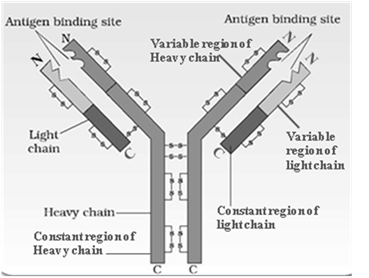
Antibodies are highly specific to specific antigens. Antibodies are produced by plasma cells (effector B-lymphocytes). The plasma cell produces about 2000 molecules of antibodies per second.
Structure of a typical Antibody:
(i) Antibody has a Y-shaped tetrapeptide molecule formed of 2 identical light chains (L chains) and 2 identical heavy chains (H chains).
(ii) The four polypeptide chains are held together by disulfide bonds (-S-S-).
(iii) Structure of Light chains:
- (a) Each light chain has a molecular weight of 25,000 Daltons.
- (b) Each light chain is made up of 214 amino acids.
- (c) The amino acid sequence of a light chain is formed of two parts
(1) Outer variable region:
consists of first 108 amino acids. It varies from one antibody to another.
(2) Inner constant region:
consists of 109th-214th amino acid and it doesn’t change.
(iv) Structure of Heavy chains:
- (a) Each heavy chain has a molecular weight of 50,000 daltons.
- (b) Each heavy chain is formed of 440 amino acids.
- (c) They are about twice as long as the light chains.
- (d) The amino acid sequence of a heavy chain is also formed of two parts:
(1) Outer variable region:
made of first 118 amino acids. It varies from one antibody to another.
(2) Inner constant region:
made of 119th to 440th amino acid. This region shows a hinge area to provide flexibility to the molecule. This hinge area allows the antibody to simultaneously bind to two epitopes that are at some distance apart on surface of antigen.
(v) Variable regions of light and heavy region constitute the antigen-binding site called paratope. It recognizes and binds to the specific antigen forming an antigen-antibody complex.
(vi) Since most antibodies carry two antigen binding sites (paratopes), they are said to be bivalent.
Antibody Fragments
The enzyme papain (derived from papaya) breaks the antibody at the hinge region to produce. A branch of immunology which deals with study of antigen-antibody interactions is called Serology.
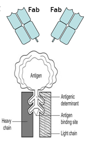
(i) Antigen and Antibody have a high level of structural complementarity between them.
(ii) The surface of antigen has antigenic determination site called epitope.
(iii) The outer surface of antibody has a variable region of amino acids called paratope.
(iv) Variations in the variable regions make each antibody highly specific for a particular antigen.
(v) The antigen-antibody bind by non-covalent bonds like hydrogen bonds and ionic bonds to produce antigen- antibody complex.
(vi) This is a highly specific epitope-paratope interaction like a lock and key mechanism.
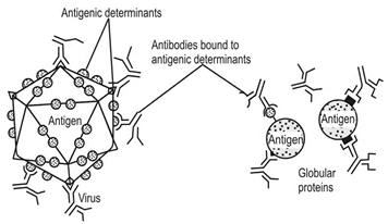
(vii) Antigen-antibody complex destroys the pathogens by the following mechanisms:
- Neutralization: It is the destruction of toxins produced by microbes.
- Opsonisation: Substances which promote cellular phagocytosis are called opsonins. Antigen-antibody complex coats the micro-organism thus making it susceptible to phagocytosis by macrophages Antibodies of the IgG type commonly acts as opsonins.
- Agglutination: It is the clumping of micro-organisms.
- Immobilisation: If antibodies are formed against antigen on the cilia or flagella of motile bacteria like Salmonella typhi, the antigen antibody reaction makes it immobile.
- Complement activation: Antigen-antibody complex causes activation of complements (plasma proteins) which have anti-microbial property.
DISEASES OF IMMUNE SYSTEM
Autoimmunity:
Autoimmunity is disease in which immune system acts against one’s own (self) cells. Lymphocytes identify self as non-self and destroy it. e.g.
|
Name of the disease |
Cells which are identified as non self |
|
Myasthenia gravis |
Skeletal muscle cells are identified as non-self and destroyed. |
|
Rheumatoid arthritis |
Synovial membrane of joints is identified as non-self and destroyed. |
|
Chronic hepatitis |
Hepatocytes are identified as non-self and destroyed. |
|
Hashimoto’s disease |
Thyroid cells are identified as non-self and destroyed. |
|
Multiple sclerosis |
Myelin sheath of neurons is identified as non-self and destroyed. |
|
Systemic Lupus Erythrematosus |
Generalized autoimmunity. |
Immunodeficiency Diseases:
Immunodeficiency means decrease in immunity. It is caused due to:
(i) Malnutrition: when there is deficiency of proteins (globulins).
(ii) Genetic Immunodeficiency:
- (a) Mutation in long arm of chromosome 20 leads to hereditary deficiency of enzyme adenosine de-aminase required for maturation of B and T cells. This causes Severe Combined Immune Deficiency (SCID).
- (b) SCID shows deficiency of both B and T cells.
- (c) SCID is the first hereditary disease to be successfully treated by gene therapy.
(iii) Acquired Immunodeficiency:
- (a) Diseases like AIDS caused by human immunodeficiency virus which reduce T- lymphocytes.
- (b) Clinical symptoms of AIDS develop when T –lymphocyte count is below
200/mm3 of blood.
- (c) Individuals with AIDS have more suppressor T cells than helper T cells
- (d) Also, according to recent studies CCR-5 (a chemokine receptor) actually opens the door knob for the entry of HIV in the blood
(1) So people with HIV and a normal CCR-5 get full blown AIDS
(2) People with defective CCR-5 move very slowly towards AIDS
(3) People without CCR-5 do not get AIDS at all.
Hypersensitivity (Allergy):
The term allergy was coined by Von Pirquet. About 10% of human population suffers from some kind of allergy.
Allergy is defined as an acquired, abnormal, hyper immune response to an agent during second or subsequent exposure. Thus for allergic reactions, body must be previously sensitized by an allergen.
Allergen: Any substance that evokes allergy is called Allergen. The common allergens are
- Food Substances like wheat, egg, milk, vegetable oils and chocolate.
- Inhalants like pollen grains, fungi, dust, smoke and perfumes.
- Contactants like chemical substances and metals.
- Infectious agents like parasites, viruses, bacteria and fungi.
- Drugs like antibiotics and aspirin.
- Physical agents like cold, heat, light and radiations.
Mechanism of allergy:
The process of allergy takes place in three steps
(1) Sensitization:
Allergen acts as a mild antigen which stimulates the formation of IgE antibodies which bind to mast cells of connective tissue. This process is called sensitization.
(2) Second stimulation:
During subsequent exposure, allergens combine with antibody-bound cells which rupture and release a large amount of histamine.
(3) Histamine reaction:
Histamine has the following effects:
- (a) Capillary dilation.
- (b) Increase in the capillary permeability. This causes leakage of body fluids.
- (c) Closure of bronchial tubes.
- (d) Increase in mucous secretions.
- (e) Pain and swelling.
During allergies, eosinophils increase in number as they have to remove antigen antibody complex. This condition is called eosinophilia.
Allergy disappears after some time as the body produces adrenaline (epinephrine) which is an anti histaminic hormone.
Types of Allergies:
The various types of Allergies are
(1) Hay Fever (allergic rhinitis):
- (a) It occurs mainly due to pollens and spores of plants.
- (b) It is characterized by increased secretion of nasal mucosa, throat-irritation, conjunctivitis.
(2) Asthma:
- (a) It occurs mainly due to house dust (containing faecal pellets of dust mites), pollen grains of plants like congress grass, pet animals, certain food and drinks, eggs, fish, red wine, drugs like aspirin, pollutants like SO2, CO2, smoke etc.
- (b) It is characterized by bronchospasm (narrowing of bronchi) and difficulty in breathing
(3) Urticaria: Allergic skin eruptions characterized by multiple, circumscribed raised pinkish itchy blisters persisting for a few days.
(4) Eczema: Dermatitis which starts with reddening of skin, formation of vesicles, rupturing of vesicles and formation of scales.
(5) Anaphylactic Shock:
- (a) It occurs due to bee-sting or some drugs like penicillin.
- (b) It is characterized by systemic allergic reaction affecting all the body tissues and can cause death.
Prevention and Treatment of Allergy:
- (i) Allergy can be prevented by identifying the specific allergen and avoiding it.
- (ii) Allergy can be treated
(1) By using antihistamine drugs like phenindramine and diphenylhydrazine.
(2) By using glucocorticoids (steroids).
Steroids reduce the immunity and thus cause relief from allergy.
Types of hypersensitivity reactions:
- (1) Immediate hypersensitivity: IgE mediated (starts within 2-30 minutes)
- (2) Antibody-dependant cytotoxic hypersensitivity:
IgG binds with self cells like platelets, neutrophils, and eosinophils etc. stimulating phagocytosis of self cells.
- (3) Immune complex mediated hypersensitivity:
Large amount of antigen-antibody complex is formed in the body and cannot be removed by reticulo-endothelial cells.e.g. serum sickness after toxoids vaccine of diphtheria
- (4) Delayed hypersensitivity:
Seen in chronic diseases like TB, fungal diseases & graft rejection. It develops in 24-72 hours.
GRAFT REJECTION:
(i) When organs like kidney, heart, lung, liver etc are grafted (transplanted) from one human being to the other, the recipient’s immune system identifies it as an antigen and starts an immune response.
(ii) Both CD4+ and CD8+ T cells participate in graft rejection.
(iii) They are responding to differences between donor and the host of their class II and class I histocompatibility molecules respectively.
(iv) If not checked, this response will lead to destruction of the graft.
GRAFT-VERSUS HOST DISEASE
(i) Red bone marrow is the site of formation of blood cells.
(ii) In some diseases, this bone marrow is affected and it needs to be replaced with a donor’s.
(iii) If there are any histocompatibility differences between the donor and recipient, the new blood cells that will be produced in the recipient is that of the donor.
(iv) The T-cells of the donor (now produced in the recipient) will elicit an immune response against the tissues of the recipient.
(v) This is called Graft versus host disease.
(vi) This can be prevented if bone marrow graft is taken from the patient himself or an identical twin
VACCINATION
History of vaccination and immunization
(i) The process of vaccination was initiated by Edward Jenner in 1790.
(ii) He observed that milkmaids did not get smallpox because they were exposed to a similar but mild form of disease called cowpox.
(iii) Edward Jenner infected the first human, James Phipps, a healthy boy of 8 years with cowpox.
(iv) Two months later, he infected the boy with smallpox.
(v) The boy did not suffer from smallpox.
(vi) Jenner suggested that an induced mild form of a disease would protect a person from a virulent form.
(vii) He thus used the term vaccine (‘vacca’= cow) and the term vaccination for protective inoculation.
(viii) Edward Jenner was the first to discover a safe and effective means to produce artificial immunity against smallpox.
(ix) Pasteur confirmed Jenner’s findings and produced vaccines for the other diseases like anthrax, rabies and chicken cholera.
(x) In 1891, a physician, Emil von Behring, infected sheep with diphtheria bacteria and withdrew blood after some time from the sheep. He separated the serum by allowing it to clot. He injected the serum into a little girl dying of diphtheria and within hours she began to recover.
Vaccines:
(i) Vaccination is the stimulation of immune system by vaccines.
(ii) Principle of vaccination (immunization) is based on memory cells.
(iii) Vaccines may be
- (a) Temporary: Cholera vaccine which lasts for 6 months.
- (b) Permanent: e.g. vaccines against whooping cough diphtheria etc.
(iv) Chemically vaccines are:
- Toxoids (toxins) like DPT vaccine against diphtheria, pertussis (whooping cough) and tetanus.
- Attenuated live pathogen (inactivated pathogen) like BCG (Bacillus Calmette Guerin) against TB, MMR (Mumps, Measles, Rubella) vaccine, OPV (oral Polio Vaccine)
- Killed pathogen like Salk vaccine (injectable) for polio, influenza vaccine, typhoid fever vaccine.
- Recombinant DNA technology is used to produce antigenic polypeptides of pathogen in bacteria or yeast. Vaccines produced using this approach allow large scale production and hence greater availability for immunization. e.g., hepatitis B vaccine produced from yeast.
Vaccines included in the Indian National Immunisation Programme are
|
No |
Name of Vaccine |
Against |
Administered |
|
1. |
BCG vaccine |
Tuberculosis |
intradermally |
|
2. |
OPV vaccine |
Polio |
orally |
|
3. |
DPT/Triple vaccine |
Diphtheria, Pertussis (whooping cough) Tetanus |
intramuscularly |
|
4. |
Measles vaccine |
Measles (Rubeola) |
intramuscularly |
ACQUIRED IMMUNE DEFICIENCY SYNDROME (AIDS):
This means deficiency of immune system, acquired during the lifetime of an individual indicating that it is not a congenital disease. ‘Syndrome’ means a group of symptoms.
History of AIDS:
(i) AIDS was first discovered in homosexuals in USA (Hatai) in 1981.
(ii) In 1983, in Paris, Luc Montagnier discovered the AIDS virus and named it
Lymphadenopathy Associated virus (LAV).
(iii) In 1984, in USA, Robert Gallo discovered the AIDS virus independently and named it
Human T- Lymphocytotrophic Virus-3 (HTLV-3).
(iv) In India, AIDS virus was first found in 10 prostitutes in Chennai in 1986.
(v) In 1986, the International Committee on Taxonomy of Virus named it Human Immunodeficiency Virus (HIV).
Causative organism of AIDS:
AIDS is caused by a virus, HIV – Human Immunodeficiency virus.
HIV belongs to the family Retroviridiae (Capable of reverse transcription)
It belongs to genus Lentivirus (Small virus causing slow viral disease) within the Retroviridiae family
Structure of HIV:
HIV is spherical and appears like a soccer ball having a diameter of 100 nm or 10-9 m.
HIV is made up of 4 parts: (1) Outer envelope (2) Inner core (3) Genetic material (4) Enzymes
(1) Outer envelope: HIV has an outer envelope made up of lipids having many knob shaped glycoprotein molecules placed at regular intervals.
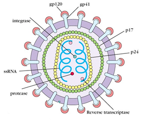
These glycoprotein molecules are of 2 types
- (i) gp 120 (surface glycoprotein)
- (ii) gp 41 (transmembrane glycoprotein)
(2) Inner core: Within the envelope, there is a protein core made of 2 coats
- (i) an outer p17 called matrix protein
- (ii) an inner p24 called capsid protein.
p24 is antigenic (capable of inducing immune response). Hence it is also called p24 antigen.
(3) Genetic material: Within the protein core there are 2 strands of RNA.
(4) Enzymes: Within the protein core there are 3 enzymes
(i) Reverse transcriptase: This is the main enzyme of HIV.
Reverse transcriptase converts the RNA of HIV into DNA within the human cell. (Conversion of RNA to DNA is called reverse transcription).
Since HIV contains reverse transcriptase, HIV is called retro virus (retro-reverse).
- (ii) Integrase helps in the integration of viral genome into the host DNA.
- (ii) Protease helps in breakdown of complex proteins formed within the host cells.
Types of HIV:
HIV is of two types:
|
No. |
HIV-1 |
HIV-2 |
|
(i) |
It is highly virulent and easily transmissible. |
It is less virulent and less transmissible. |
|
(ii) |
It is more common and is seen in India (Asia), America and Europe |
It is seen in Africa. |
|
(iii) |
It is further categorized into 4 groups M, N, O & P |
It is further categorized into 8 groups A- H. |
|
(iv) |
About 90% of AIDS cases in the world are caused by HIV-1 group M. |
How did human beings get HIV?
(i) The African green monkey contains Simian Immunodeficiency Virus which is very similar to HIV.
(ii) It is believed that this monkey bit or scratched some human beings.
(iii) Thus the virus spread to humans through the wounds.
(iv) Some people believe that in 1950’s the kidney of the African monkey was used to prepare a polio vaccine.
(v) This vaccine was given to 3,25,000 African children and maybe this vaccine is caused the spread of SIV to humans. SIV further mutated to form HIV.
Source of infection of HIV
HIV is present in large quantity in blood, semen, pre-seminal fluid which comes out before ejaculation, vaginal fluid and C.S.F. of infected person.
HIV is also present in smaller quantity in tears, saliva, breast milk and urine of infected person.
Modes of transmission of AIDS
AIDS spreads by par-enteral route (par-besides/beyond enteral-digestive system).
It spreads are follows:
(i) By intimate sexual contact: HIV spreads by sexual contact with an infected person.
The sexual contact could be
- (a) Homosexual contact: Sexual contact between individuals of the same sex.
- (b) Heterosexual contact: Sexual contact between individuals of the opposite sex.
- (c) With multiple sexual partners:
About 80% cases of AIDS are spread by sexual contact.
- (d) It could also spread through oral or anal sex.
(ii) From infected blood:
HIV spreads by contact with infected blood. Infected blood can spread:
- (a) During blood transfusion
- (b) From infected instruments, needles and syringes during surgery, acupuncture, tattooing, ear piercing, drug addiction etc.
- (c) Sharing infected razors and toothbrushes.
(iii) During procedures: HIV may also spread during certain procedures like
- (a) Organ transplantation.
- (b) Dialysis (procedure done during kidney failure in which waste products are removed from circulating blood).
- (c) Artificial insemination using infected semen.
(iv) From infected mother to child (Transplacental transmission):
- (a) HIV may also spread from mother to child through placenta during labour (delivery).
- (b) Spread of infection from mother to child is called vertical / transplacental transmission.
- (c) A nursing mother can transmit HIV to her baby from her breast milk.
- (v) AIDS cannot be spread by insect bite due to lysosome present in the saliva of insects.
Mechanism of action of HIV
HIV infects only those cells that have CD4 receptors. CD4 receptors are present on
(1) helper T lymphocytes (in blood)
(2) alveolar macrophages (in lungs)
(3) microglia (neuroglia in central nervous system).
(4) The virus can also infect other lymphoid cells such as B cells and lymphoid cells of the brain and testes.
After getting into the body of the person, the virus enters into macrophages where RNA genome of the virus replicates to form viral DNA with the help of the enzyme reverse transcriptase.
This viral DNA gets incorporated into host cell’s DNA and directs the infected cells to produce virus particles.
The macrophages continue to produce virus and in this way acts like a HIV factory. Simultaneously, HIV enters into helper T-lymphocytes, replicates and produce progeny viruses. The progeny viruses released in the blood attack other helper T-lymphocytes leading to a progressive decrease in the number of helper T-lymphocytes in the body of the infected person.
During this period, the person suffers from bouts of fever, diarrhoea and weight loss. Due to decrease in the number of helper T lymphocytes, the person starts suffering from infections that could have been otherwise overcome such as those due to bacteria especially Mycobacterium, viruses, fungi and even parasites like Toxoplasma. The patient becomes so immuno-deficient that he/she is unable to protect himself/herself against these infection
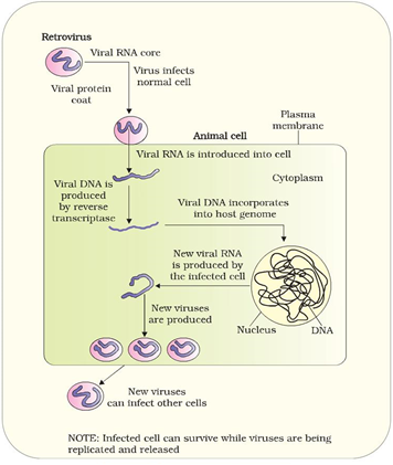
Incubation period of AIDS:
(i) It is the period between the infection (i.e. entry of causative agent) and the appearance of the first symptom of disease.
(ii) Incubation period of AIDS is 5-10 years.
(iii) During this period the patient is HIV+ but doesn’t suffer from AIDS. AIDS is last stage of disease
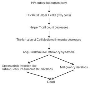
Clinical features of AIDS.
(i) Initial infection stage: It starts 2-3 weeks after HIV infection.
Only 3-5% of those newly infected have mononucleosis-like symptoms that may include fever, chills, aches, swollen lymph glands, and an itchy rash.
These symptoms disappear, and there are no other symptoms for 9 months or longer.
(ii) Asymptomatic carrier stage:
It continues for 5-9 years and during this phase the patient is HIV positive but has not developed AIDS. In this phase the individual is infected with HIV but does not show any symptom hence it’s called asymptomatic phase. Although the individual exhibits no symptoms during this stage, he or she is highly infectious. In this phase, it is very difficult to say whether the person has AIDS or no. Therefore, it is also called blind phase.
The standard HIV blood test for the presence of antibody becomes positive during this stage.
(iii) Early Symptomatic HIV infection (AIDS Related Complex):
Major Signs:
- Weight Loss more than 10% of body weight. Due to excessive loss of body weight, this disease in also called Slim disease or Wasting syndrome.
- Persistent Diarrhoea for more than 1 month.
- Persistent Fever for more than 1 month
Minor Signs:
(1) Lymphadenopathy (swelling of lymph nodes): The patient has enlarged lymph nodes at least 1cm in diameter all over the body especially in the neck, armpits and groin that persist for 3 months or more.
(2) Persistent Cough for more than 1 month. Excessive coughing is due to lung infections like tuberculosis (bacterial) and Pneumocystic carinii pneumonia (fungal).
(3) Oropharyngeal Infection like Candidiasis is an infection of mouth and throat caused by a fungus Candida albicans. It causes curdy white coating on tongue.
(4) Skin Infection: like Herpes zoster (shingles).
(5) Night sweats: Excessive sweating at night.
Full blown AIDS stage:
(1) Helper T cell count (CD4 count) reduces below 200mm3 of blood.
(2) Severe Opportunistic infections: These infections are called opportunistic because the body can usually prevent them with normal immunity but due to an immunocompromised state in AIDS, they can easily occur. These infections include infections with Mycobacterium, Viruses, fungi & even parasites like Toxoplasma.
(3) Thrombocytopenic purpurea: Decreased count of blood platelets.
(4) Meningitis, partial paralysis.
(5) The following cancers (malignancies) develop:
- (i) Kaposi sarcoma: It is the cancer of inner lining of blood vessels. It is thought to be caused by Human Herpes virus 8.
- (ii) Lymphoma: It is the cancer of lymph nodes.
(6) Dementia (Loss of memory): HIV kills brain cells, resulting in memory loss, inability to think clearly, loss of judgment and/or depression.
(7) Finally death occurs. Few patients are called Non-progressors as in them AIDS develop very slowly or never at all due to a genetic difference that prevents the virus from damaging their immune system, only an impaired immune system gives them the opportunity to get started
Diagnosis of AIDS
AIDS can be detected by the following tests:
(i) E.L.I.S.A. (Enzyme Linked Immuno Sorbent Assay)- Screening test.
This test is used to detect antibodies produced by the human body against HIV.
The principal enzyme used in ELISA is alkaline phosphatase prepared using horse radish.
After entry of HIV in the body, a period of 3-6 months is required by the human body to produce the antibodies. During this period ELISA test will be negative.
The period when the person is infected by HIV but ELISA in negative is called window period. If ELISA is negative, either the patient does not have disease or may be in the window period. If ELISA is positive, the person may or may not have HIV.
ELISA test may shows a positive result in some other conditions like Leprosy, T.B., Malaria, Cancer, Hepatitis B, Rheumatoid arthritis, Haemophilia, Pregnancy, Kidney failure, Flu, etc.
(ii) Western blot test -Confirmatory test for ELISA positive cases
Western Blot is more specific and more reliable for HIV but it is expensive. It is also detects the antibodies against HIV. This test also has a window period of 3-6 months
(iii) PCR (Polymerase chain reaction) to detect of viral genome.
Polymerase chain reaction (PCR) doesn’t have window period. It is the fastest test for HIV.
Treatment of AIDS
AIDS is a fatal viral disease without cure. Drugs can only give temporary relief and prolong life. These drugs do not destroy the HIV but only stop its multiplication and growth in human body by inhibiting the important enzymes which are necessary for viral replication.
Hence these drugs are called viristatic drugs. They are as follows:
(i) Reverse transcriptase inhibitors:
Reverse transcriptase inhibitors bind to enzyme reverse transcriptase and prevent connection of RNA to DNA. They are:
- AZT (Azidothymidine or Zidovudine) is the most popular drug against HIV
- Nevirapine: Nevirapine alone or in combination with zidovudine is normally advised by the doctors to HIV the pregnant women to ensure that their babies do not get infected.
- DDI (Dideoxyinosine)
(ii) Protease inhibitors: Protease inhibitors bind to enzyme protease and inhibit its action. eg. Foscarnet, Indinavir.
(iii) Integrase inhibitors: Integrase inhibitors bind to enzyme Integrase and inhibit its action. eg. Raltegravir
(iv) Interferon enhancement drugs: Interferons are antiviral substances and are used in viral diseases. Ribavirin is a drug which enhances the action of interferons and increases the number of T helper cells.
A combination of several anti-retro viral drugs called HAART-highly active Anti-Retroviral Therapy has been very effective in reducing the viral load no. of HIV particles blood stream.
AIDS Awareness programmes:
India started National AIDS Control Programme in 1987 by National AIDS Control Organization (NACO)
WHO started various programmes to create awareness. 1st Dec is declared as world AIDS day.
CANCER
It is uncontrolled unorganized and abnormal growth of cells, forming a mass or group of undifferentiated malignant cells called neoplasm (tumour). Cancer has come from the Greek word “Karkinos’ and Latin word ‘cancrum’ which means crab. Just like a crab does not easily leave a place where it sticks, cancer does not easily leave the organ which it affects. It is also called Karka Rog. The scientific study of cancer is called Oncology.
Properties of Cancer:
(1) Abnormal cell division:
Cancer occurs only in those cells which undergo cell division. Hence cancer never occurs in cardiac cells and neurons. Nucleus is very important for cell division. Hence cancer can never occur in enucleated cells like RBCs and platelets. Cancer cells don’t enter G0 stage (resting phase) of cell cycle. Cancer cells contain an enzyme telomerase which is absent in normal cells. Telomerase induces unlimited cell divisions. Therefore cancer cells are immortal.
(2) Cancer cells show: Anaplasia- abnormal structure and Hyperplasia- increase in number
(3) Cancer cells are highly abnormal cells with an
- Abnormal cell membrane The cell membrane is abnormal in shape because the cytoskeleton becomes thin. The cell surface has many spicules (spines) like a sea urchin, to shield themselves from the defensive immune cells of the body. Glycoproteins and glycolipids present on plasma membrane are lost or modified.
- Abnormal nucleus: Nucleus contain more amount of DNA. Therefore the nucleus is large with ucleolus. Nucleus stains darker than normal, hence called hyperchromatic nuclei.
(c) Abnormal cytoplasm
The cells organelles in the cytoplasm show the following abnormalities:
- The mitochondria are swollen.
- Ribosomes and endoplasmic reticulum are more in number.
- Microtubules and microfilaments in the cytoplasm tend to disorganize.
(d) Abnormal functions
- Cancer cells respire anaerobically.
- They compete with normal cells for nourishment, hence act as parasites.
- Cancer cells induce nearby cells into forming nutrition-bearing blood vessels and growth promoting chemicals. This property of cancer cells of forming
new blood vessels is called angiogenesis.
(e) Loss of contact inhibition:
Normal body cells exhibit a property termed contact inhibition which prevents the uncontrolled growth and division of cells. Cancer cells fail to exhibit contact inhibition hence continue to divide giving rise to masses of cells called neoplasm or tumor.
(f) Metastasis (Spread): The spread of cancerous cells to distant sites is termed metastasis.
A cancerous cell manages to move throughout the body using the blood or lymph systems, destroying healthy tissue in a process called invasion.
Genetic basis of development of cancer
Humans have two types of genes:
- Oncogenes: Oncogenes are cancer producing genes. About 100 different types of oncogenes have been identified.
- Anti-oncogenes: Genes which oppose the production of cancers are called anti-oncogenes or cancer suppressing genes or tumour suppressor genes.
- eg (a) MTS-1: multiple tumour suppressor
- (b) nM-23: non-metastatic gene
- (c) p53 gene: Dr. Harris proved that tiny alterations in p53 gene leads to lung cancer. In p53 - p stands for protein and 53 is the molecular mass in kilodaltons.
Tumour suppressor genes are also called guardian of genome.
(iii) In a healthy individual,
- Oncogenes are present in an inactive form called proto-oncogene or c-onc
- Anti-oncogenes are present in an active form.
Thus development of cancer is prevented in normal cells.
(iv) Whenever a carcinogen enters the body, it alters the DNA of cells in such a way that
- Inactive oncogenes (proto-oncogenes) are activated.
- There is damage to anti-oncogenes making them defective.
Thus cancer is developed.
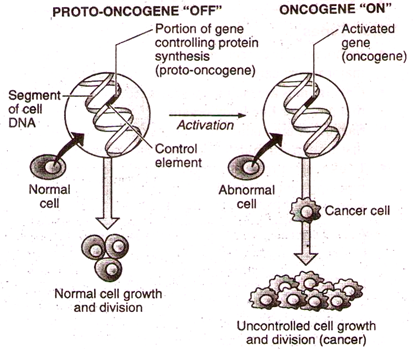
Cancer development in Immuno-comprised state like AIDS
Normally the lymphocytes (i.e. the immune system) destroy cancer cells. If the lymphocytes becomes weak (i.e. the immune system fails) like in AIDS or if the person is given immune suppressant drugs, the cancer can develop easily.
Causes of Cancer (Carcinogenic agents)
|
No |
Agents |
Carcinogens |
Cancer caused |
|
1. |
Physical agents |
Ultraviolet rays |
Skin cancer (in white skin) |
|
X-rays |
Leukemia (in radiologist) |
||
|
Wearing tight saree (clothing) |
Skin cancer |
||
|
Khangri is a bag containing burning coal tied around the abdomen worn by kashmiris to keep themselves warm. |
Khangri cancer (abdominal skin cancer) |
||
|
Radium watch factory workers paint luminous dials with radium. |
Bone cancer |
||
|
Radioactive isotopes and ionizing radiations released by atomic bomb |
Leukemia and other cancers seen in Japanese |
||
|
2. |
Chemical agents |
Tobacco smoke and coal tar (soot) contains benzopyrene and nitrosamines |
Oral and lung cancer |
|
Vinyl chloride |
Liver cancer |
||
|
Cadmium oxide |
Prostate cancer |
||
|
Aromatic amines eg. 2-naphtylamine, 4-aminobiphenyl |
Urinary bladder cancer |
||
|
Asbestos, arsenic, aniline dyes, chromium, nickel and mustard gas |
Lung cancer |
||
|
Benzene |
Bone marrow cancer |
||
|
Diethylstilbestrol (DES) |
Cancer of vagina |
||
|
Increased testosterone in body |
Prostate cancer |
||
|
Increased estrogen in body |
Breast and vaginal cancer |
||
|
3. |
Biological agents Virus causing cancer are called onco-virus. Baltimore, Dulbecco and Temin proved that viruses cause cancer by changing gene structure. They received Nobel Prize in 1975. |
Hepatitis B virus (HBV) |
Liver cancer (Hepatoma) |
|
Human papilloma virus (HPV), Epstein Barr virus, Herpes simplex type-2. |
Cervix cancer |
||
|
Human immunodefiency cirus (HIV) |
Kaposi sarcoma |
||
|
Epstein-Barr virus |
Burkitt’s lymphoma (jaw tumor) and nasopharyngeal cancer |
||
|
Aflatoxin (a toxin produced by a fungus Aspergillus flavus) |
Liver cancer |
||
|
Schistosoma haematobium (parasite) |
Urinary bladder cancer |
||
|
4. |
Mechanical agents |
Mucosal irritation due to chewing tobacco/ betel nut |
Lip and mouth cancer |
|
Mucosal irritation due to broken tooth |
Tongue cancer |
||
|
5. |
Nutritional agents |
Alcohol (caused 3% of all cancer deaths) |
Cancer of entire digestive system, liver and pancreas |
|
Smoked fish |
Stomach cancer |
||
|
Beef |
Intestinal cancer |
||
|
Beer |
Rectal cancer |
||
|
High fat diet |
Breast cancer |
||
| - | - |
Synthetic colours and synthetic salts like Monosodium glutamate (Aginomoto) used in food are also found to be cancerous. High protein rich diet, high carbohydrate food like potato chips and French fries release carcinogenic agents, on heating which may cause intestinal cancer. |
|
Clinical features of cancer
Clinical features are also called danger signals of cancer or red signals or warning signs of cancer.
American Cancer Society has given seven danger signals of cancer as follows
- Change in bowel or bladder habit.
- A scab, sore or ulcer that fails to heal within 3 weeks.
- Unusual bleeding or discharge through natural orifices like nipple, urethra, nose, vagina etc.
- Thickening or mass in the breast or other body parts.
- Indigestion or difficulty in swallowing.
- Obvious change in a wart, blemish or a mole (enlargement, bleeding or itching)
- Nagging cough or hoarseness.
- Excessive loss of body weight without apparent cause.
- Unexplained low grade fever.
Types of Cancer Based on their spread
On the basis of their spread tumours are of 2 types:
|
Benign Tumour |
Malignant Tumour |
||
|
1 |
Metastasis (spread of cancer) does not occur. |
1 |
Metastasis (spread of cancer) occurs |
|
2 |
It is covered by capsule. |
2 |
It is not covered by capsule. |
|
3 |
It is round/oval in shape. |
3 |
It is irregular in shape. |
|
4 |
Cells more adhesive. |
4 |
Cells less adhesive. |
|
5 |
Tumour is non invasive. |
5 |
Tumour is invasive. |
|
6 |
Tumour cells are well differentiated |
6 |
Tumour cells are undifferentiated. |
|
7 |
It grows slowly. |
7 |
It grows rapidly. |
|
8 |
Benign tumour does not cause pain. |
8 |
Malignant tumour causes pain. |
|
9 |
Tumour can be excised surgically. |
9 |
Tumour can not be cured by surgery. |
|
10 |
Mortality is generally low. (except brain tumours which are dangerous as they can press against vital areas) |
10 |
Mortality-high. |
Types of Cancer based on their location:
On the basis of their location cancer is of the following types :
- Sarcoma: The cancer of mesodermal organs is called sarcoma. eg. Cancer of muscle (sarcoma), bone (osteoma), cartilage, fats and connective tissue.
- Carcinoma: The cancer of epithelium (covering internal and external parts of the body) eg. cancer of breast, lungs, intestine, colon, skin, liver, pancreas, cervix, kidney etc
- Leukaemia: The cancer of blood (WBCs) is called leukaemia. Cancerous Bone marrow cells produce excess of WBCs, so WBC count increases.
- Leukemia is of two types:
- Myeloid leukemia Cancer of granulocytes (basophils, eosinophils and neutrophils).
- Lymphoid leukemia Cancer of lymphocytes.
- Lymphoma: The cancer of lymph node, spleen and tissues of immune system They produce excess of lymphocytes.eg. Hodgkin’s lymphoma.
- Adenomas: The cancer of glandular tissue is called Adenoma. eg. Cancer of thyroid gland, pituitary gland, adrenal gland etc.
- Melanoma: Carcinoma of melanocytes (skin).
- Lipoma: Tumour (generally benign) of adipose tissue.
Commonest Cancers in the world
- Commonest global cancer in males: lung cancer
- Commonest Indian cancer in males: oral cancer
- Commonest global cancer in females: breast cancer
- Commonest Indian cancer in females: cervix cancer
- Commonest age group of cancer: 40-60 years
Staging of Cancer
The International Union Against Cancer (IUAC) has evolved the “TNM” classification of cancer.
- Stage 1: T-represents primary tumour size.
- Stage 2: N-represents involvement of lymph nodes.
- Stage 3: M-represents metastasis or distant spread.
Diagnosis/Investigations of Cancer:
(1) Biopsy: A small piece of affected organ is cut into thin sections, stained and examined under microscope (histopathological studies) by a pathologist.
(2) FNAC (Fine Needle Aspiration Cytology):
A needle is introduced into the affected organ and some cells of the organ are aspirated. (ie ; removed by suction).These cells are then examined under the microscope.
(3) Endoscopy:
- (a) Gastroscopy is used to detect stomach cancer (helps in internal visualization of stomach).
- (b) Laproscopy is used to detect cancer of pelvic and abdominal organs.
(4) Tumour markers:
Some chemicals present on the surface of cancer cells are used to detect cancers. These chemicals are called tumour markers.
(5) CT Scan: Computerized Axial Tomography uses X-Rays to generate a three dimensional image of the internal organs
(6) MRI: Magnetic Resonance Imaging uses strong magnetic fields and non-ionizing radiation to detect pathological and physiological changes in the living tissues including cancers of internal organs.
(7) Blood: Done to detect blood cancers.
(8) DNA probe analysis: Done to detect different cancers.
(9) Antibodies for cancer specific antigen are also used.
(10) Molecular Biology techniques are applied to detect genes in individuals with inherited susceptibility to certain cancers. Identification of such genes, which predispose an individual to certain cancers, may be very helpful in prevention of cancers. Such individuals may be advised to avoid exposure to particular carcinogens to which they are susceptible (e.g., tobacco smoke in case of lung cancer)
(11) Screening for specific cancers:
- Cancer of cervix: The method of detection is called Papanicolaou test or pap test. In this test a cervical smear is examined.
- Screening for lung cancer: Chest radiography and sputum cytology are done for screening of lung cancer.
- Screening for breast Cancer:
- Self breast examination
- Palpation by a physician
- Mammography
- Thermography
- Prostate cancer: Prostate specific antigen (PSA) tests for prostate carcinoma.
- Intestinal cancer: Occult blood tests (hidden blood is detected) for intestinal cancers
Treatment of Cancer
- Surgery: Tumor is cut and removed.
- Radiotherapy: Radiation given in a fixed dose kills rapidly dividing cancer cells.eg. Cobalt 60, X-rays, I131
- Brachytherapy: It is an advanced radio-therapeutical technique in which a very high dose of radiation is given to small volume of body tissues in short period from small radioactive sources like Radium, Cobalt 60 etc.
- Chemotherapy: Specific chemotherapeutic drugs are administered that check cell division by inhibiting DNA synthesis. They kill cancerous cells but have side effects like hair loss, anemia etc.
eg: Vinca rosea (Catharanthus roseus) is source of two anticancer drugs; Vinblastine and Vineristine for leukemia treatment. Vinca rosea is also known as sada bahar/sada phuli.
Some other anticancer drugs used are:
- Mercaptopurine
- 6-Aminopterin
- Photoferin is used for treatment of throat cancer. It is prepared from cow’s blood.
- Alkylating agents (cell poisons) like Nitrogen mustard, Alkyl sulfonates, Cyclophosphamide, Triethylene, thiophosphamide etc.
- Antimetabolites like Folic acid antagonists, purine antagonists
- Antibiotics like Actinomycin D (attacks DNA of cancer cells).
- Combination therapy: It involves treatment by combination of surgery, radiation therapy & chemotherapy.
- Interleukin-2: Interleukins are signaling molecules released by WBCs themselves. They stimulate other WBC’s (lymphocytes) to kill the cancer cells.
- Hormonal therapy: Cancer caused by one hormone is treated by another hormone which has a neutralizing effect (Opposite Effect). Eg : In females, high estrogen (female sex hormone) level caused breast cancer. Hence, Testosterone (Male sex hormone) is used for treatment of breast cancer. Anti breast cancer drugs are Tamoxifen, Taxol which counter-act the effect of estrogens.
- Controlling angiogenesis: Folkman, a chemical substance obtained from a fungus prevents blood vessel formation and thus, prevents rapid growth of tumour.
- Vaccine (under trial): A vaccine is being used for treatment of Melanoma (tumour of melanocytes of skin) and hepatoma (tumour of liver).
- Interferon: These are antiviral substances used in treatment of cancer caused by viruses.
- Use of Monoclonal antibodies: Monoclonal antibodies are taken from hybridoma cells. (Hybridoma = cell formed by the fusion of single plasma cells with tumor cell).
- Bone marrow transplantation: Recommended in case of leukaemia
- Immunotherapy:It is the recent approach to treat cancer. Here, the natural anticancer immunological defense mechanism is supplemented to kill cancer cells.
ADOLESCENCE:
Adolescence is the period accelerated physical and mental growth extending between childhood and adulthood.
Characteristics of Adolescence:
- Adolescence extends from puberty to complete sexual maturation i.e period between 12-18 years
- There is an accelerated physical growth with development of reproductive organs.
- There are physical and physiological changes like increase in height and weight, reduction in fat (puppy fat) muscle distribution and development, glandular secretions and sexual characteristics. There is a spurt in physical growth, which may be as much as 7.5-15 cm in a single year.
- Congnitive changes include the processes of intellectual growth and all the mental processes involved in thinking. The ability to think abstractly and systematically develops during adolescence.
- Ability to reason about moral, ethical and religious issues emerges.
- There is a growing appreciation about other people’s motives, feelings and social roles. There is a search for identity.
- Adolescents consider themselves more idealistic and less materialistic, and are skeptical about the efficiency and ethics of government, business and other social institutions.
- Values such as love, friendship, privacy, tolerance and self-expression are emphasized.
Common Problems of Adolescence:
- Acne: It is most common problem of adolescents of both the sexes.
It is characterized by appearance of small sized blackheads on the skin especially that on the face. It occurs due to hormone androgen. It occurs due to overproduction of sebum from the sebaceous (oil) glands of the skin and blockage of skin pores.
- Hypochondria: It refers to an excessive preoccupation or worry about having a serious illness. It is also called health phobia.
It is a psychosomatic disorder in which the adolescents suffer from anxiety and undue concern about their health.
- Anorexia nervosa: It is the inability to consume food in some adolescents due to fear of obesity & disfigurement of body. This disorder is commonly seen in young women who try to lose body weight by resorting to voluntary food restrictions or dieting.
This causes many complications such as weakness, anemia & irregularities in menstrual cycle. Bullimia nervosa It involves cycles of excessive eating and purging by self induced vomiting.
- Neurasthenia: It denotes a condition with symptoms of fatigue, anxiety, headache, impotence, depressed mood and insomnia (inability to sleep).It is the result of exhaustion of the central nervous system’s energy reserves. Americans are supposedly more prone to neurasthenia, which resulted in its nickname Americanitis. It is associated with upper class individuals in sedentary employment.
- Phobias: A phobia refers to repulsive behavior and intense fear from certain things or creatures.
Some of the common phobias are
- Algophobia: Fear of pain.
- Nyctophobia: Fear of darkness.
- Xenophobia: Fear of strangers.
- Claustrophobia: Fear of enclosed spaces.
- Phasmophobia: Fear of ghosts.
- Social awkwardness and exhibitionism, and aggressive self assertion.
- Physiological disorders: In some females, physiological disorders like absence of monthly periods are seen.
- Post-traumatic stress disorders: It may occur due to traumatic experience like rape, robbery, certain accidents etc.
- Addiction to drugs, alcohol, smoking, etc., is also very common among the adolescents.
- Juvenile delinquency: Adolescents may often indulge in crime and violence. It is called juvenile delinquency
SMOKING/ TOBACCO ADDICTION
Burning tobacco in the form of bidi, cigarette, pipe etc. and inhaling tobacco smoke is called smoking Tobacco is derived from the dried leaves of plant
(1) Nicotiana tabacum which is a native of America and
(2) Nicotiana rustica which is a native of Europe.
The dried tobacco leaves are either smoked or chewed with lime or snuffed.
Tobacco smoking was started by Red Indians of America about 400 years ago. In 16th century, tobacco smoking spread to Europe and other parts of the world.
Contents of Tobacco smoke:
Tobacco smoke contains:
- Nicotine is the chief, toxic, alkaloid substance in tobacco. Nicotine is the chief addictive substance of tobacco. Nicotine stimulates the nervous system and gives pleasure. It relaxes muscles. It stimulates adrenal medulla to secrete adrenaline constricting blood vessels. This increases heart rate and blood pressure.
In high doses, nicotine damages neurons as it causes overstimulation of neurons. If Nicotine present in one cigarette (60 mg) is injected intravenously, it may prove fatal.
- Tar particles are particulates (i.e. minute unburnt black particles) present in tobacco smoke. It contains polycylic aromatic hydrocarbons like benzopyrene, nitrosoamines, methyl chloranthrene etc. They are chief carcinogenic substances of tobacco.
- Gases like carbon monoxide, CO2, H2S, nitrogen oxides, NH3 etc. While smoking, nearly 10% of the smoke is inhaled, remaining is dispersed in air.
Adverse effects of Smoking
- Reduction in oxygen carrying capacity of Haemoglobin: Tobacco smoke contains carbon monoxide which combines irreversibly with haemoglobin to form carboxy haemoglobin. Hence oxygen carrying capacity of blood decreases. This can cause hypoxia (decreased oxygen supply) to brain, heart and other organs. Due to this there is mental dullness, dizziness, headache, breathlessness, coma and even death
This is the basic cause of heart disease in heavy smokers. The formation of carboxy haemoglobin may even lead to congestive heart failure
- Effect on respiratory system:
(a) Bronchitis:
Tobacco smoke irritates epithelium of trachea, bronchi and alveoli of lungs. This irritation of epithelium produces more and more secretions. The excessive secretions are removed by coughing. Thus there is continuous coughing especially in the morning, called smoker’s cough.
(b) Emphysema:
It’s a chronic obstructive lung disease characterized by loss of elasticity of lung tissue, destruction of alveoli supporting structures and destruction of capillaries feeding alveoli.
Tar particles present in tobacco smoke destroy macrophages which protect the respiratory tract. They also cause paralysis of cilia and hence the secretions and micro-organisms cannot escape from the respiratory system.
Since alveoli are damaged, exchange of CO2 and O2 decreases leading to breathlessness.
- Effects on circulatory system:
(a) Hypertension and heart attack:
Tobacco smoking causes increase of adrenalin secretion which increases blood pressure, heart beat rate by constricting the arteries. Nicotine damages the bicuspid valve (mitral valve) of the heart.
(b) Buerger’s disease includes
- Constriction of small arteries.
- Arteriosclerosis : loss of elasticity of arteries.
- Thrombosis : clotting inside blood vessels.
All these lead to reduced blood supply to organs. If there is complete blockage of blood supply to an organ, there is gangrene (death of tissue). The gangrenous part has to be amputated.
- Effects on digestive system:
Smoking increases the secretion of gastric juice containing HCl which can cause:
- Gastritis is the irritation of epithelial lining the stomach.
- Ulcers in stomach and duodenum causing abdominal pain, nausea, vomiting, loss of appetite and bleeding from stomach.
- Cancer: Polycylic aromatic hydrocarbons like benzopyrene, in tobacco smoke, are carcinogenic. They cause cancers like lung cancer (bronchogenic carcinoma), oral cancer and throat cancer. Benzopyrene causes mutation of p53 gene (Anti-oncogene) leading to lung cancer.
Oral cancer is common in reverse smokers (Smoking with burning end in mouth) commonly seen in Andhra Pradesh.
Cancer related statistics:
- 33% of all cancers are caused by tobacco.
- In males, 50% of all cancers are caused by tobacco.
- In females, 25% of all cancers are caused by tobacco.
- 85-95% lung cancers are due to smoking.
- Amblyopia: Smoking of strong tobacco causes Amblyopia (i.e. Dim Vision).
- Effect on Immune System: Smoking reduces immunity of the body.
- Effects on pregnancy: Smoking during pregnancy leads to foetal growth retardation and even Abortion.
Control Measures of smoking
- Use of mass media (like TV, radio, newspaper) to create awareness about ill-effects of smoking.
- Every year 31st May is observed as No Tobacco Day to create awareness.
- Psychotherapy: Counseling & motivating a patient to generate a strong will power to quit smoking.
- Substitution therapy: Substitution of smoking by ayurvedic cigarettes (without nicotine) and nicotine patches & gums.
ALCOHOLISM
Non-medical, regular and excessive use of alcohol leading to complete physical and psychological dependence is called alcoholism. The addict is called alcoholic.
Alcohol is a colourless fluid produced by the fermentation of carbohydrates with the help of yeast enzymes. It has some positive aspects hence used in various medicines.
Alcohol beverages with low concentration of alcohol (5-15%) are beer, cider, wine etc. Alcohol beverages with high concentration of alcohol (40-50%) are whisky, brandy, rum, gin, vodka etc. The active ingredient of alcoholic beverages is ethyl alcohol.
Metabolism of alcohol in the body:
- Absorption: Alcohol is absorbed from stomach (20%) and upper part of intestine (80%) and transported to the cells of the body. There is no digestion of alcohol prior to absorption. From here, the alcohol reaches the liver, as the cells of the body cannot utilize alcohol.
- Oxidation: In the liver cells, alcohol is oxidized to form acetaldehyde and acetic acid with the help of enzyme acetaldehyde dehydrogenase. A large amount of heat energy is produced. As a result, blood circulation to the skin increases, making the skin warm.
- Excretion: Small amount of alcohol escapes oxidation. Lung excretes little alcohol in exhaled air, so the breath smells of alcohol. Kidney excretes little alcohol in urine. Skin excretes little alcohol in sweat.
Endorphin Theory of alcohol addiction
The cause of Alcohol addiction has been explained by the endorphin theory as follows:
Endorphin is a morphine like chemical substance, naturally present in brain. It reduces tension & stress. In some people endorphin is less, and hence they suffer from stress. They drink alcohol to overcome it. Alcohol further decreases the endorphin level of the brain and the person becomes more dependent on alcohol.
Adverse Effects of Alcoholism
- Acute symptoms (immediate effects): Alcohol causes vasodilation and increases oxidation rate, that produces lot of heat giving a person a pleasant warm feeling. This is called alcoholic flush.
It also causes mental confusion and dizziness, slurred speech, blurred/doubled vision, staggering gait and defective muscular co-ordination. There is loss of judgment and self control, loss of emotional control (i.e he laughs or weeps foolishly), loss of moral sense (i.e. antisocial behaviour) and even loss of consciousness.
- Effect on the nervous system: Alcohol is a CNS depressant (Actually, alcohol has a biphasic effect on CNS. In lower concentrations it acts as stimulant but at higher levels alcohol is a depressant.
Alcohol acts as Sedative (i.e. alcohol produces sleep), Analgesic (i.e. alcohol relieves pain). Anaesthetic (i.e. alcohol causes loss of sensation). Alcohol causes inflammation of Neurons (Neuritis).It can cause permanent damage to neurons Long term consumption of Alcohol causes loss of memory due to brain damage (Amnesia).
- Effects on the liver: Due to repeated detoxification in the liver alcohol may cause toxic hepatitis.
Alcohol also causes deposition of fat in the form of tawny yellow coloured hard nodules in the liver. This stage is called fatty Liver. This is reversible if alcohol consumption is stopped. If alcohol is not stopped, the liver cells are damaged. and gradually the liver shrinks in size and hardens. This is called cirrhosis of liver.
Also, liver fails to perform its function i.e. liver failure. Chronic alcoholism may cause liver cancer. In the presence of alcohol, the cytochrome P-450 enzyme produces carcinogens from body chemicals.
- Effects on digestive system: Excess and concentrated alcohol consumption causes,
- Gastritis is the irritation of epithelium lining the stomach. A pain killer drug called aspirin also causes gastritis. Hence when taken along with alcohol, it causes severe damage to gastric mucosa.
- Ulcer in stomach and duodenum causes abdominal pain, nausea, vomitting, loss of appetite and bleeding from stomach.
- Cardiomyopathy : Alcohol also affects working of the heart and increases chances of heart attack by increasing blood pressure. Due to high blood pressure cardiac muscles may undergo hypertrophy. The heart muscles become rigid and lose flexibility and the heart may be enlarged. This condition is called cardiomyopathy.
- Cancer: Alcoholism causes cancer of liver, oesophagus and stomach.
- Nutritional deficiency: An alcoholic does not take care of his diet. Hence, he suffers from deficiencies of carbohydrate, proteins, vitamins and minerals.
Thiamine (Vitamin B1) deficiency in alcoholism leads to Wernicke’s encephalopathy (Acute phase) in which there is changes in mental state, unsteady gait due to reduced muscular co-ordination and ocular disturbances like double vision.
It is followed by Korsakoff’s Syndrome (Chronic phase) in which there is, paralysis of hands and feet, mental confusion, loss of memory and hallucinations.
- Foetal alcohol syndrome: It is developed in babies of mothers who have consumed alcohol heavily during pregnancy. Baby is under weight, mentally retarded or may have deformed organs.
- Effect on Kidney: Long term consumption of alcohol causes kidney failure. Hence, blood urea level increases.
- Effects on Urine formation: Alcohol decreases secretion of ADH causing increased urine output but urine is hypertonic. Also, in alcoholics, along with heat, large amount of water is also given out, but through the skin. So, water level in the body decreases. Therefore, even though alcohol decreases secretion of ADH, urine output is not increased to a large extent as the amount of water available in the body itself is less. Thus, Kidney effectively produces small amount of concentrated urine i.e. Oliguria and Hypertonic Urine
- Withdrawal syndrome/delirium tremens (DT): If a chronic alcoholic stops/reduces alcohol consumption, he suffers from,
- Illusions and hallucinations (visual/auditory).
- Anxiety, fear and restlessness.
- Increase in heart rate and blood pressure.
- Loss of sleep and appetite.
- Convulsions (rum fits). They last for 3 to 10 days.
- Hypoglycaemia: Alcohol decreases blood sugar level
- Tunnel Vision: Alcohol affects the eyes leading to decrease in field of vision.
- Acidosis: Ethyl alcohol gets detoxified in the liver to form acetic acid. Also there may be impurities in alcohol like methyl alcohol which gets detoxified in the liver to form formic acid.
This causes excessive accumulation of acids in blood leading to decrease in pH. Acidosis leads to respiratory failure (stoppage of respiration).
- Hangover Effect: An intoxicated person may suffer from headache, drowsiness etc. even after he has regained his senses. This effect is called a hangover.
One should not drive after consumption of alcohol because
- Alcohol affects judgment.
- Alcohol affects coordination: Coordination of the limbs, the head and the eyes are impaired affecting the driver’s control of the car.
- Alcohol affects alertness: A driver becomes less watchful and fails to observe objects outside his vehicle.
- Alcohol affects vision: Vision becomes blurred and unsteady. Often the field of vision is reduced (Tunnel Vision).
- Alcohol increases reaction time: The driver takes more time to react to unexpected situations, e.g., a child running across a street.
- Alcohol affects behaviour: Intoxicated drivers become rash, careless and erratic. They tend to speed and take risks
Control measures of alcoholism
- Medical Measures:
- Chlorodiazepoxide prevents withdrawal symptoms.
- Aversion Therapy/Antabuse Therapy:
- Disulfiram tablet is given to the patient. Now, if the patient drinks even small amount of alcohol, he suffers from nausea, vomiting, headache, flushing, palpitation, mental confusion etc. Hence, he avoids alcohol.
- Emetine hydrochloride injection is given to patient. Now, if the patient sees, smells or drinks alcohol, he starts vomiting. Hence, he starts hating alcohol. (Emetine hydrochloride is toxic, hence it is not used any more).
- Psycho Social Measures:
- Psychotherapy: Counseling of an addict to quit alcohol.
- Social Therapy (Family Therapy): The family member and friends should be counseled and should encourage alcoholics to quit alcohol.
- Occupational Therapy: Rehabilitation of alcoholic after de-addiction.
- Alcoholic Anonymous: It is an international, voluntary organization where ex-alcoholics share their personal experiences with alcoholics and motivate them to quit alcohol.
