Fertilization
Fertilization refers to fusion of male and female gametes (i.e. spermatozoon and ovum). It generally takes place in the middle segment at ampullary-isthmus junction of the fallopian tube. It involves following events:
Transport of Gametes
Before fertilization, the ovum and sperms reach the ampulla for fertilization. Fertilisation can only occur if the ovum and sperms are transported simultaneously to the ampullary–isthmic junction. This is one of the reason why not all copulations lead to fertilisation and pregnancy.
Transport of ovum. At the time of ovulation the secondary oocyte is directly expelled into the peritoneal cavity and then enters into the fallopian tube.
Mechanism
The fimbriae of the fallopian tube are internally lined by ciliated epithelium. When ovulation occurs, the fimbriae of infundibulum encircle the surface of ovary, and pick up the ovum and then direct it towards the ostium by continuous beating of cilia. The contraction of smooth muscle fibres present in the wall of fallopian tube also help in transport of oocyte. These contractions are increased at the time of ovulation under the influence of oestrogen.
- Structure of oocyte: The released secondary oocyte consists of 23 unpaired chromosomes surrounded by the inner membranous layer called zona pellucida consisting of glycoproteins and on its outer side surrounded by corona radiata consisting of granulosa cells arranged in multiple layers. These cells are held together by matrix composed of hyaluronic acid.
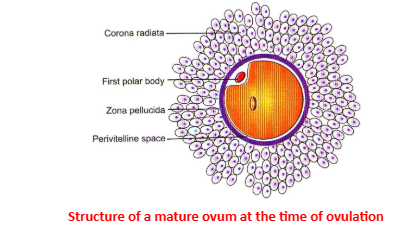
- Here zona pellucida is also known as primary egg membrane and corona radiata is the secondary egg membrane.
- Fate of secondary oocyte It is held up at ampulla-isthmic junction for 24 to 72 hours. During this period if viable sperm penetrates it, then fertilization takes place leading on to pregnancy. On the other hand, if fertilization does not occur, the oocyte dies out and degenerates.
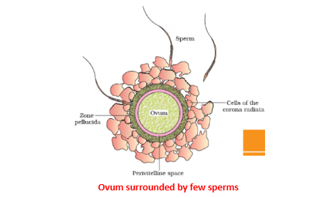
- Transport of sperms in the female genital tract. After ejaculation, several million sperms (average – 200 million sperms per ejaculation) get deposited in the vagina. Out of these about 100-150 sperms manage to reach on to ovum and only one is able to penetrate it. This is because of several factors that affect the transportation and accessibility of the sperm to ovum.
Sperm capacitation
Freshly ejaculated sperm are unable or poorly able to fertilize. Rather, they must first undergo a series of changes known collectively as capacitation. Capacitation is associated with removal of adherent seminal plasma proteins, reorganization of plasma membrane lipids and proteins. It also seems to involve an influx of extracellular calcium, increase in cyclic AMP, and decrease in intracellular pH.
Capacitation occurs while sperm reside in the female reproductive tract for a period of time, as they normally do during gamete transport. The length of time required varies with species, but usually requires several hours. The sperm of many mammals, including humans, can also be capacitated by incubation in certain fertilization media (In vitro method).
Sperm that have undergone capacitation are said to become hyperactivated, and among other things, display hyperactivated motility. Most importantly however, capacitation appears to destabilize the sperm's membrane to prepare it for the acrosome reaction.
Sperm-Zona pellucida binding
Binding of sperm to the zona pellucida is a receptor-ligand interaction with a high degree of species specificity. The carbohydrate groups on the zona pellucida glycoproteins (ZP3) function as sperm receptors.
The acrosome reaction
The acrosome reaction provides the sperm with an enzymatic drill to get through the zona pellucida. The same zona pellucida protein that serves as a sperm receptor also stimulates a series of events that lead to many areas of fusion between the plasma membrane and outer acrosomal membrane. Membrane fusion (actually an exocytosis) and vesiculation expose the acrosomal contents, leading to leakage of acrosomal enzymes from the sperm's head.
As the acrosome reaction progresses and the sperm passes through the zona pellucida, more and more of the plasma membrane and acrosomal contents are lost. By the time the sperm traverses the zona pellucida, the entire anterior surface of its head, down to the inner acrosomal membrane, is denuded. Sperm that lose their acrosomes before encountering the oocyte are unable to bind to the zona pellucida and thereby unable to fertilize.
Penetration of the Zona Pellucida
The constant propulsive force from the sperm's flagellating tail, in combination with acrosomal enzymes, allow the sperm to create a tract through the zona pellucida. These two factors - motility and zona-digesting enzymes- allow the sperm to traverse the zona pellucida.
Sperm-Oocyte Binding
Once a sperm penetrates the zona pellucida, it binds to and fuses with the plasma membrane of the oocyte. Binding occurs at the posterior (post-acrosomal) region of the sperm head. Sperm glycoprotein called fertilin, which binds to a protein in the oocyte plasma membrane and may also induce fusion.
Egg Activation and the Cortical Reaction
Prior to fertilization, the egg is in a quiescent state, arrested in metaphase of the second meiotic division. Upon binding of a sperm, the egg rapidly undergoes a number of metabolic and physical changes that collectively are called egg activation. Prominent effects include a rise in the intracellular concentration of calcium, completion of the second meiotic division and the so-called cortical reaction.
The cortical reaction refers to a massive exocytosis of cortical granules seen shortly after sperm-oocyte fusion. Cortical granules contain a mixture of enzymes, including several proteases, which diffuse into the zona pellucida following exocytosis from the egg. These proteases alter the structure of the zona pellucida, inducing what is known as the zona reaction. Components of cortical granules may also interact with the oocyte plasma membrane.
The Zona Reaction
The zona reaction refers to an alteration in the structure of the zona pellucida catalyzed by proteases from cortical granules. The critical importance of the zona reaction is that it represents the majorblock to polyspermy in most mammals. This effect is the result of two measurable changes induced in the zona pellucida:
- The zona pellucida hardens. Crudely put, this is analogous to the setting of concrete. Runner-up sperm that have not finished traversing the zona pellucida by the time the hardening occurs are stopped in their tracks.
- Sperm receptors in the zona pellucida are destroyed. Therefore, any sperm that have not yet bound to the zona pellucida will no longer be able to bind, let alone fertilize the egg.
Post-fertilization Events
Following fusion of the fertilizing sperm with the oocyte, the sperm head is incorporated into the egg cytoplasm. The nuclear envelope of the sperm disperses, and the chromatin rapidly loosens from its tightly packed state in a process called decondensation. In vertebrates, other sperm components, including mitochondria, are degraded rather than incorporated into the embryo.
Chromatin from both the sperm and egg are soon encapsulated in a nuclear membrane, forming pronuclei.
Each pronucleus contains a haploid genome. They migrate together, their membranes break down, and the two genomes condense into chromosomes, thereby reconstituting a diploid zygote.
First Week of Development
First week of development begins immediately after fertilization. Changes seen during this week include:
- Cleavage of zygote
- Formation of morula, transportation to uterine cavity and conversion into blastocyst, and
- Implantation of blastocyst.
Cleavage of zygote (0 to 3 days)
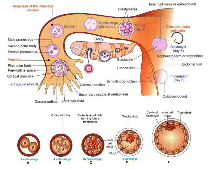
∙The fertilized and activated ovum (zygote) is much larger (140 μm) than an average cell and starts dividing immediately. The process of repeated mitotic division of the zygote within the zona pellucida, resulting in rapid increase in the number of cells, is called cleavage. The cells formed are called blastomeres. In humans and other mammals, due to scanty deutoplasm there occurs holoblastic or total cleavage.
∙Ordinarily, cleavage divisions occurs in quick succession without any intermittent growth or synthetic phase; they are rapid, rhythmic and repeated mitotic divisions. In mammals, including humans, cleavage divisions are among the slowest in the animal kingdom. Also, the cleavage divisions are asynchronous from the very beginning; the number of resultant cells increase, following arithmetic progression.
Site and duration of cleavage. The cleavage occurs in the uterine tube upto 3 days after fertilization.
Cleavage in Humans
First cleavage division occurs 30 hours after fertilization and two blastomeres are formed. Second cleavage division occurs 10 hours after first and four blastomeres are formed. Third cleavage division is completed by end of third day w.r.t fertilization.
Formation of morula, transportation in uterine cavity and its conversion into blastocyst.
Morula – At about sixteen cells-stage, the blastomeres tightly align themselves against each other to forms a compact ball of cells called morula (mulberry). The process of compaction leads to segregation of inner cells (which form inner cell mass) from the surrounding cells (which form the external cell mass).
Transportation of morula into the uterine cavity then occurs. The transportation is assisted by the fluid currents and ciliary movements of epithelial cells of the fallopian tube and uterus.
Formation of Blastocyst
In the cavity of uterus, the fluid from the lumen of uterus (uterine milk) seeps through the zona pellucida and outer-cell-mass of the morula. With continued accumulation of fluid the morula is converted into blastocyst. The blastocyst, thus consists of:
Zona pellucida is the outer covering
- Embryoblast, is a group of clustered blastomeres, formed from inner-cell mass of the morula. It is attached to one pole of the blastocyst and later give rise to tissues of the embryo proper and some extraembryonic tissues.
- Trophoblast is a thin outer layer of cells formed from the outer-cell-mass of the morula. It contributes to extraembryonic structures and embryonic part of placenta.
- Blastocoel is the fluid filled space inside the blastocyst.
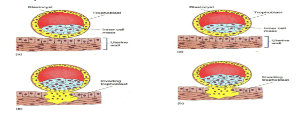
Implantation (Nidation)
Endometrium of uterus is meanwhile converted into decidua by enlargement of stromal cells which become vacuolated and filled with glycogen and lipids.
- The blastocyst then erodes and burrows into the decidua with the help of proteolytic enzymes secreted by the trophoblast.
- Blastocyst grows deeper in the decidua progressively till it is completely buried under the mucosa. The implantation or embedding of the blastocyst takes place on 6th or 7th day after fertilization. This type of implantation is also known as interstitial implantation.
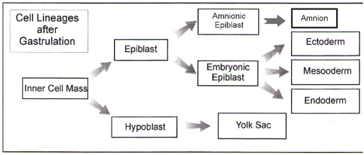
Epiblast and Hypoblast
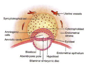
- On 8th day after ovulation the embryoblast is converted into a bilaminar disc consisting of following two layers.
- Epiblast, the upper layer, is thick and composed of regularly arranged columnar cells. It is continuous at the periphery with amnion
- Hypoblast, the lower layer, is thin and made of irregularly arranged polyhedral cells. It is continuous at its periphery with the yolk sac.
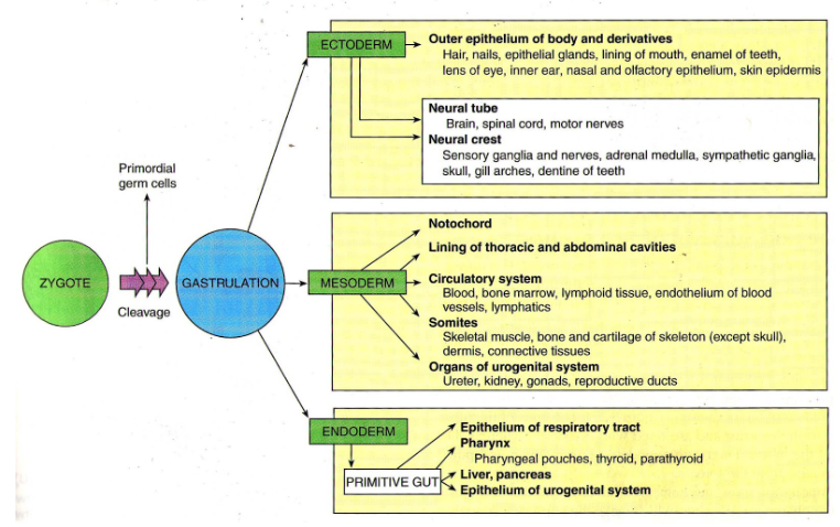
- The blastocyst then undergoes gastrulation to produce the three primary germinal layers. This involves cell movements that eventually help to attain new shape and morphology of the embryo. These cell movements are called morphogenetic movements.
- Following gastrulation, the embryo passes through the phase of neurulation, during which the primordium of nervous system, the neural plate, is laid down. Gradually, the organ rudiments appear and the process of organogenesis is inaugurated. During organogenesis, the various organs of the foetus are established and they attain functional state.
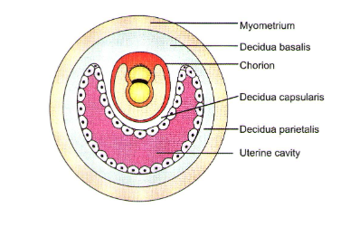
Decidua
It refers to the changed endometrium which converts it into decidual cells. Due to effect of increasing progesterone levels in maternal blood, the stromal cells of endometrium get converted into decidual cells by becoming enlarged, vacuolated and filled with glycogen and lipids. The stored glycogen and lipid are the source of nutrition for the embryo till placenta takes up this function.
Parts of decidua are named as below
- Decidua basalis or decidual plate refers to the part where maternal part of placenta is to develop.
- Decidua capsularis is that part of the decidua which overlies the embryo and separates it form the uterine cavity.
- Decidua parietalis refers to the part of the decidua lining the rest of uterine cavity.
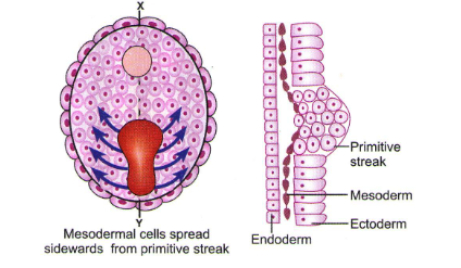
Gastrulation
As described above, the embryonic disc at the end of second week is a bilaminar disc consisting of epiblast and hypoblast. At the beginning of third week there occurs, the most characteristic event, the gastrulation, which establishes all the three germ cells, changing the embryonic disc from a bilaminar to a trilaminar structure. During this period, the embryo may be referred to as a gastrula.
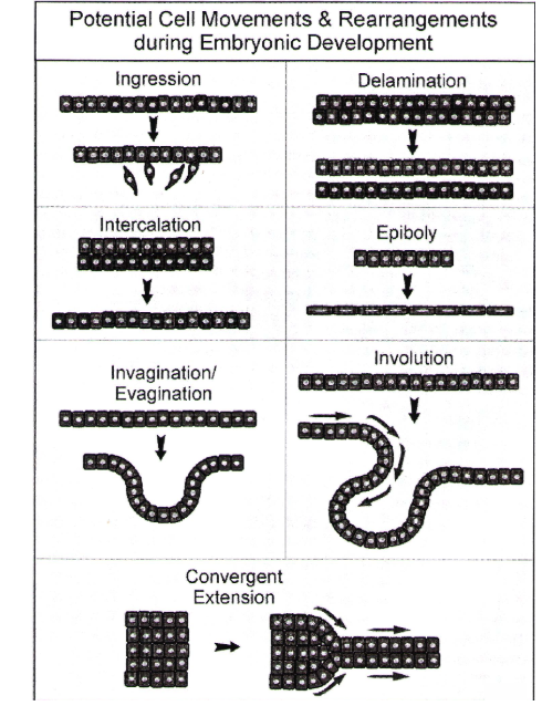
Cell Movements & Rearrangements during Embryonic Development
During human gastrulation, cells move over the blastodisc surface, enter the primitive streak and move internally. On the surface they move in association with other cells but once they turn the corner around the lip of the primitive streak the cells separate as individuals to migrate internally to form the mesoderm and endoderm. Following are some of the types of cell movements that occur in animal embryos.
- Ingression: cells break away from the tissue and migrate as individuals
- Delamination: layers of cells separate from each others more or less as sheets of cells
- Intercalation: two cell layers interlace with each other
- Epiboly: a form of cell spreading in which cells flatten out; this allows them to cover a much larger surface area.
- Invagination (Evagination): a tissue layer folds in (out)
- Involution:cells move over a lip of tissue and into the interior
- Convergent Extension: cells reorganize to form less layers allowing the cells to extend out from a point.
Fake Maps of Gems Layers
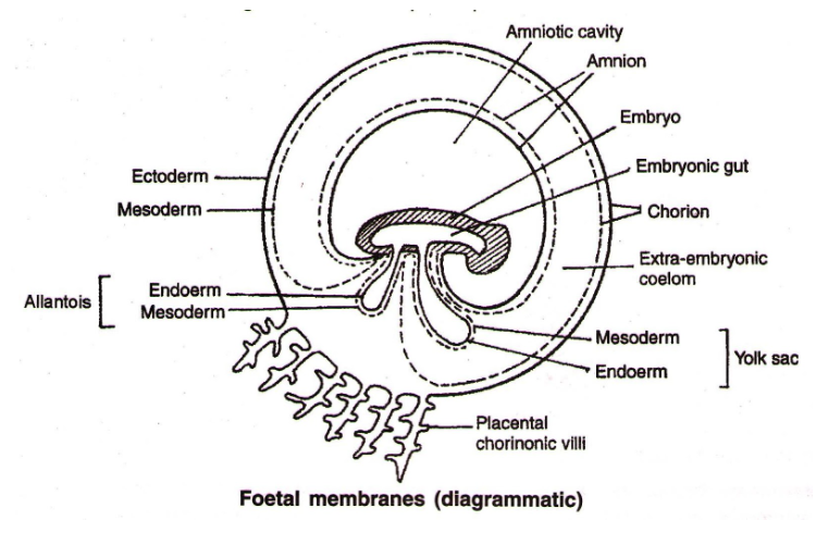
Extra Embryonic Membranes
The extraembryonic or foetal membranes are the amnion, chorion, allantois and yolk sac. Amnion provides a fluid medium to the developing embryo; it prevents desiccation of the embryo and function as a shock absorber. As the human egg is devoid of yolk, the yolk sac develops as an evolutionary process. It is very small and gradually degenerates and shrinks. The chorion and allantois take part in the formation of placenta in most of the mammals. So in mammals it is called chorioallantoic placenta. In humans it is chorionic placenta.
Placenta
Placenta is a temporary foetomaternal connection formed during pregnancy. It forms an important circulatory link between the mother and foetus. It is the only tissue in the body which has cells from two different individuals of two different generations.
Formation of Placenta
- The placenta consists of two major portions:
- Maternal part of placenta, (derived from deciduas), and
- Fetal part of placenta (derived from chorion)
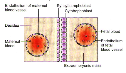
- Placental membrane consists (from foetal side) of following layers
- Endothelium and basement membrane,
- Surrounding mesenchymal tissue (connective tissue),
- Syncytiotrophoblast
- The foetal placenta grows intimacy and invades the uterine mucosa with its chorionic villi. The degree of intimacy is so strong that eventually the blood vessels of the villi are literally bathed in the mother’s blood. This becomes possible due to erosion of the uterine mucosa, including its epithelium, connective tissue and the endothelial lining. This type of placenta is known as haemochorial placenta.
Types Of Placentea
On the basis of the structure which fuses with the chorion the placenta in mammals is categorized into two types.
|
Placenta |
Characteristic |
Example |
|
Vitellochorion placenta or (Choriovitelline placenta) |
It is formed by yolk sac and chorion, i.e. Vitellochorion membrance. Allantois in such placenta is reduced |
Marsupials or Metatherian (e.g. Kangaroo – Macropus) |
|
Allantochorion placenta |
Such placenta is formed jointly by allantois and chorion. Yolk sac is reduced. |
Eutherian mammals. |
On the basis of distribution of villi.
|
Placenta |
Characteristic |
Example |
|
Diffused placenta |
The villi are diffused all over the surface of the embryo |
Horse and Pig |
|
Coteledonary placenta |
Villi are present over the embryo in the form of patches/groups called cotyledons |
Cattle |
|
Zonary placenta |
Villi are present in the form of a belt around the embryo |
Carnivorous mammals (Dog) |
|
Discoidal placenta |
Villi are present in the form of a disc. It can be monodiscoidal or bidiscoidal |
Rodent and Lagomorphs (Rabbit) |
|
Metadiscoidal placenta |
Villi in the beginning are distributed all over the surface but are later restricted only to a disc shaped area |
Primates (Monkeys. apes and Human) |
On the basis of Histology
The name of the placenta is assigned after the name of the maternal tissue which is in direct contact of foetal tissue.
|
Placenta |
Characteristic |
Example |
|
Epitheliochorial placenta |
It is the simplest placenta The number of barriers is six (3+3) The uterine epithelium comes in contact of chorionic epithelium, hence the same epitheliochorial. |
Horse, pig |
|
Placenta |
Characteristic |
Example |
|
Syndesmochorial placenta |
The number of barriers is five (2-from material side and 3-from foetal side) The connective tissue of uterus comes in contact of chorionic epithelium. Epithelial lining of uterus is eroded. |
Cattle |
|
Endotheliochorial placenta |
The number of barriers of four (1 + 3) The endothelium of material blood vessels comes in contact of chorionic epithelium. The maternal connective tissue is also eroded due to deep penetration of chorionic villi |
Carnivorous mammals (dogs, cats) |
|
Haemochorial placenta |
The number of barriers is three (0 + 3) The blood of maternal sides comes in contact with chorionic epithelium, hence name haemochorial. The endothelial lining also erodes due to more branching. |
Primates |
|
Haemoendotheliochorial placenta |
Number of barriers is one – i.e. the endothelial lining of foetal blood vessels only Blood of mother side is in contact of endothelial lining of chorionic blood vessels. Chorionic epithelium and connective tissue of foetus also erode. |
Rabbit |
Functions of Placenta
- The placenta acts as an ultrafilterate. Soluble inorganic and organic materials, nutrients, hormones, antibodies against diphtheria, small pox, scarlet fever, measles, etc. can pass from the mother to the foetus.
- It also helps in the exchange of gases between the mother and the foetus. The placenta also helps in the elimination of metabolic wastes of the foetus.
- Placenta act as an endocrine gland and synthesizes large quantities some hormones, such as human chorionic gonadotropin (HCG), chorinoic thyrotropin, chorionic corticotrophin, chorionic somatomammotropin, estrogens and progesterone. The HCG stimulates the corpus luteum of pregnancy that continues to secrete progesterone until the end of pregnancy.
- In addition, it secretes relaxin that facilitates parturition by softening the connective tissue of the symphysis pubis. The metabolic activity of the placenta is almost as great as that of the foetus itself. The umbilical cord connects the foetus to the placenta.
Developmental stage of foetus during different months of gestation
- In human beings, after one month of pregnancy, the embryo’s heart is formed. The first sign of growing foetus may be noticed by listening to the heart sound carefully through the stethoscope.
- By the end of the second month of pregnancy, the foetus develops limbs and digits.
- By the end of 12 weeks (first trimester), most of the major organ systems are formed, for example, the limbs and external genital organs are well-developed.
- The first movements of the foetus and appearance of hair on the head are usually observed during the fifth month.
- By the end of 24 weeks (second trimester), the body is covered with fine hair, eye-lids separate, and eyelashes are formed.
- By the end of nine months of pregnancy, the foetus is fully developed and is ready for delivery.
How to calculate the probable date of birth:
- From date of fertilization. Expected date of delivery is most accurately indicated as 266 days or 38 weeks after fertilization. However, usually it is not possible to know the exact date of fertilization.
- From date of last menstrual period. Practically, most obstetricians calculate the Expected date of delivery as 280 days or 40 weeks after the first day of last menstrual period.
Parturition
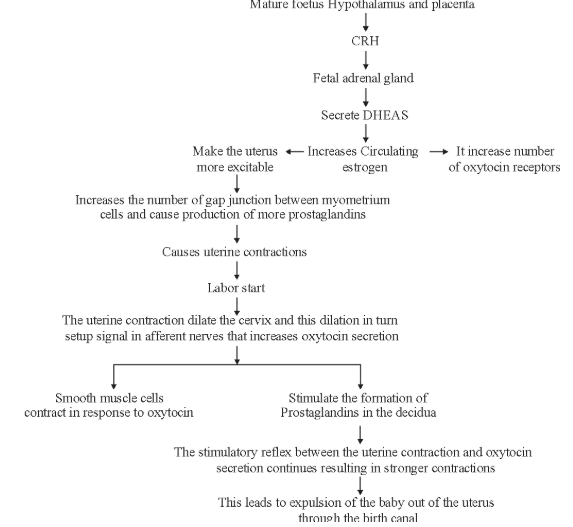
Parturition is induced by a complex neuroendocrine mechanism. The signals for parturition originate from the fully developed fetus and the placenta which induce mild uterine contractions called foetal ejection reflex.
Lactation
Following childbirth and the expulsion of the placenta, the maternal blood concentration of placental hormones decline rapidly, and the action of prolactin is no longer inhibited.
Prolactin stimulates the mammary glands to secrete large quantities of milk. This hormonal effect does not occur until two or three days following birth, and in the meantime, the glands secrete a thin, watery fluid called colostrum. Although colostrum is relatively rich in proteins, particularly protective antibodies (IgA), its concentrations of carbohydrates and fats are lower the those of milk.
Cleavage
- Holoblastic-equal type and the pattern is biradial, indeterminate type.
- Takes place during passage of zygote through fallopian tube to uterus.
- During 2nd cleavage division one of the two blastomeres divide little later resulting in 3-cell stage for a short while. But, soon typical 4-cell stage appears and subsequently morula is formed.
- In many mammals zona pellucida remains intact housing the morula.
- Developing embryo acquires nutrition from the uterine fluid/blood of mother from the very beginning.
Cleavage of zygote (0 to 3 days)
- The fertilized and activated ovum (zygote) is much larger (140 μm) than an average cell and starts dividing immediately. The process of repeated mitotic division of the zygote within the zona pellucida, resulting in rapid increase in the number of cells, is called cleavage. The cells formed are called blastomeres. In humans and other mammals, due to scanty deutoplasm there occurs holoblastic or total cleavage.
- ∙rdinarily, cleavage divisions occurs in quick succession without any intermittent growth or synthetic phase; they are rapid, rhythmic and repeated mitotic divisions. In mammals, including humans, cleavage divisions are among the slowest in the animal kingdom. Also, the cleavage divisions are asynchronous from the very beginning; the number of resultant cells increase, following arithmetic progression.
Site and duration of cleavage
The cleavage occurs in the uterine tube upto 3 days after fertilization.
Cleavage in humans
First cleavage division occurs 30 hours after fertilization and two blastomeres are formed. Second cleavage division occurs 10 hours after first and four blastomeres are formed. Third cleavage division is completed by end of third day w.r.t fertilization.
Types of Cleavage
- Holoblastic cleavage – The cleavage furrow passes through whole of the egg
|
Cleavage |
Characteristic |
Examples |
|
Equal holoblastic cleavage |
equal sized cells are produced. |
Microlecithal eggs. |
|
Unequal holoblastic cleavage |
unequal sized cells are produced. |
Mesolecithal eggs |
- Meroblastic cleavage – The cleavage furrow do not pass through the entire egg, but only a part of the egg undergoes segmentation. Such cleavage is also known as partial cleavage.
|
Cleavage |
Characteristic |
Examples |
|
Discoidal cleavage |
Cleavage occur in a disc shaped portion of the cytoplasm (Blastodisc) |
In Reptiles and birds eggs |
|
Superficial cleavage |
Cleavage occur on the surface of the eggs and the centre (yolky part) remains unsegmented. |
Insect eggs |
On the basis of the ‘fate of the cells’ the cleavages can be categorized into –
|
Cleavage |
Characteristic |
Examples |
|
Indeterminate cleavage |
The fate of the cells in early cleavage is not determined, and if the cells at 2 or 4 celled-stage are separated, each can form a complete embryo. The animals having indeterminate cleavage can produce identical twins. |
In eggs of Echinoderms and Chordates (Deuterostomes) |
|
Determinate cleavage |
The fate of the cells in early cleavage is fixed or determined. If even a single cell, in a 4-celled stage is damaged, the embryo will be defective, because the structures, to be formed by the damaged cell, will not develop. The animals with such cleavage can not produce identical twins. |
In eggs of Nematodes, annelids and mollusks (Protostomes) |
The cleavage can also be classified on the basis of arrangement (pattern) of the cells in early embryos.
|
Cleavage |
Characteristic |
Examples |
|
Radial cleavage |
The cells of upper and lower tier lie in the same vertical axis or at the same radius. |
In eggs of Deuterostomes |
|
Spiral cleavage |
The cells of one tier lie at the junction of the cells of the other tier. This means the cells of different tier are spirally arranged. Sometimes, in radially arranged cells, the cells of upper tier rotate in clock or anti clock wise manner to assume spiral pattern. The eggs which show Indeterminate cleavage or Radial pattern are called Regulative eggs, and the eggs which show determinate cleavage of spiral pattern are called Mosaic eggs. |
Protostomes |
Planes of Cleavage
|
Planes of Cleavage |
Characteristic |
|
Meridional plane |
When a cleavage furrow passes from animal pole to vegetal pole through the centre of the egg, thus dividing the egg into two equal halves, the plane of cleavage is called meridional. |
|
Vertical plane |
If the above plane does not pass through the centre of the egg and unequal sized cells are produced, the plane is called vertical. |
|
Equatorial plane |
The cleavage furrow passes transversely through the centre of the egg and equal sized cells are produced. |
|
Latitudinal plane |
The cleavage furrow is transverse but does not pass through the centre of the egg and the cells produced are unequal in size. |
|
Tangential plane |
Cleavage occurs at the surface of the egg and very unequal size cells are produced. |
Childbirth Occurs in Three Stages
Illustrates the three stages of childbirth, dilation, expulsion, and placental.
Stage 1, the dilation stage, gets its name from the dilation of the cervix. This phase begins when uterine contractions start and generally lasts 6–12 hours; but it can last much longer.
Stage 2, the expulsion stage, begins after the cervix is dilated to 10 centimeters and the baby is engaged. At this time, uterine contractions usually occur every 2 or 3 minutes and last 1–1.5 minutes each.
Stage 3 :
The final stage of delivery is the placental stage. It results in the expulsion of the placenta. The placenta remains attached to the uterine wall for a short while. It is then expelled by uterine contractions, usually within 15 minutes of childbirth.
Changes in baby
- Expansion of lung and start of breathing.
- Closure of ductus aorticus and foramen ovale.
- Adult pattern of blood flow through heart, aorta and pulmonary arteries.
- In some babies these switch over may not be completed due to inadequate synthesis of nitric oxide.
Secretion of Milk
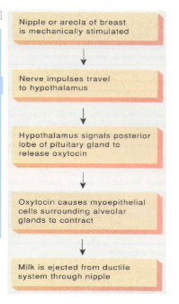
- 3-4 days after delivery breasts begin to secrete milk in sufficient amount, stimulated by prolactin.
- Its release from the teats is stimulated by oxytocin or due to suckling stimulus by baby.
- Milk contains an inhibitory peptide. If breasts are not fully emptied peptides accumulate and inhibit further production of milk.
- The first milk, colostrum, secreted just after the child birth contains lots of proteins and antibodies that provide passive immunity to the newborn.
Egg Membranes
The eggs in sponges and most of the coelenterates are naked, i.e., they have no coatings outside the plasma membrane. But in most of the other animals there are covering outside the plasma membrane, called egg membranes. These egg membranes are of three types.
|
Egg Membranes |
Characteristic |
Examples |
|
Primary egg membranes |
∙They develop mainly from primary oocyte ∙They are formed before ovulation when the egg is in the ovary |
Vitellinle membrane and zona pellucida. |
|
Secondary egg membranes |
They are formed from follicle cells. They are formed before ovulation when the egg is in the ovary. |
Corona radiata (mammals), chorion (insects) |
|
Tertiary egg membranes |
They are formed/secreted from the structures other than the ovary. They are formed after ovulation when the egg is in oviduct or uterus etc. |
Shell, Shell membranes and Albumen (in bird’s eggs), and jelly (in frog’s eggs) |
Type 0f Eggs
- On the bases of amount of yolk
|
Type of Eggs |
Characteristic |
Examples |
|
Microlecithal/Oligolecithal eggs |
A very small amount of yolk (as compared to the amount of Cytoplasm) present |
Marsupials and Eutherian mammals, echinoderms and cephalochordates. |
|
Mesolecithal eggs |
A moderate amount of yolk is present. In frog the yolk is concentrated towards vegetal pole. |
Cyclostomes and amphibians (frog) |
|
Macrolecithal/megalecithal/ Polylecithal eggs |
A huge amount of yolk is present |
Insects, fishes, lizards, birds and prototherian mammals. |
- On the bases of distribution of yolk
|
Type of Eggs |
Characteristic |
Examples |
|
Homolecithal/Isoleithal eggs |
Yolk is sparse and is thinly or uniformly distributed throughout the cytoplasm. (The cytoplasm is homogeneous). |
Metatherians, Eutherian mammals, Echinoderms and Cephalochordates |
|
Centrolecithal eggs |
The yolk is concentrated towards the centre of the egg and is surrounded by cytoplasm all around. |
Insects |
|
Telolecithal eggs |
Yolk is concentrated at one end (vegetal pole) of the egg. |
Cyclostomes, Fishes, Amphibians, Reptiles, Birds and Prototherian mammals. |
Childbirth Occurs in Three Stages
Illustrates the three stages of childbirth, dilation, expulsion, and placental.
Stage 1, the dilation stage, gets its name from the dilation of the cervix. This phase begins when uterine contractions start and generally lasts 6–12 hours; but it can last much longer.
Stage 2, the expulsion stage, begins after the cervix is dilated to 10 centimeters and the baby is engaged. At this time, uterine contractions usually occur every 2 or 3 minutes and last 1–1.5 minutes each.
Stage 3 :
The final stage of delivery is the placental stage. It results in the expulsion of the placenta. The placenta remains attached to the uterine wall for a short while. It is then expelled by uterine contractions, usually within 15 minutes of childbirth.
Changes in baby
Expansion of lung and start of breathing.
Closure of ductus aorticus and foramen ovale.
Adult pattern of blood flow through heart, aorta and pulmonary arteries.
In some babies these switch over may not be completed due to inadequate synthesis of nitric oxide.
Secretion of Milk
3-4 days after delivery breasts begin to secrete milk in sufficient amount, stimulated by prolactin.
Its release from the teats is stimulated by oxytocin or due to suckling stimulus by baby.
Milk contains an inhibitory peptide. If breasts are not fully emptied peptides accumulate and inhibit further production of milk.
The first milk, colostrum, secreted just after the child birth contains lots of proteins and antibodies that provide passive immunity to the newborn.
Egg Membranes
The eggs in sponges and most of the coelenterates are naked, i.e., they have no coatings outside the plasma membrane. But in most of the other animals there are covering outside the plasma membrane, called egg membranes. These egg membranes are of three types.
|
Egg Membranes |
Characteristic |
Examples |
|
Primary egg membranes |
∙They develop mainly from primary oocyte ∙They are formed before ovulation when the egg is in the ovary |
Vitellinle membrane and zona pellucida. |
|
Secondary egg membranes |
They are formed from follicle cells. They are formed before ovulation when the egg is in the ovary. |
Corona radiata (mammals), chorion (insects) |
|
Tertiary egg membranes |
They are formed/secreted from the structures other than the ovary. They are formed after ovulation when the egg is in oviduct or uterus etc. |
Shell, Shell membranes and Albumen (in bird’s eggs), and jelly (in frog’s eggs) |
Type 0f Eggs
- On the bases of amount of yolk
|
Type of Eggs |
Characteristic |
Examples |
|
Microlecithal/Oligolecithal eggs |
A very small amount of yolk (as compared to the amount of Cytoplasm) present |
Marsupials and Eutherian mammals, echinoderms and cephalochordates. |
|
Mesolecithal eggs |
A moderate amount of yolk is present. In frog the yolk is concentrated towards vegetal pole. |
Cyclostomes and amphibians (frog) |
|
Macrolecithal/megalecithal/ Polylecithal eggs |
A huge amount of yolk is present |
Insects, fishes, lizards, birds and prototherian mammals. |
- On the bases of distribution of yolk
|
Type of Eggs |
Characteristic |
Examples |
|
Homolecithal/Isoleithal eggs |
Yolk is sparse and is thinly or uniformly distributed throughout the cytoplasm. (The cytoplasm is homogeneous). |
Metatherians, Eutherian mammals, Echinoderms and Cephalochordates |
|
Centrolecithal eggs |
The yolk is concentrated towards the centre of the egg and is surrounded by cytoplasm all around. |
Insects |
|
Telolecithal eggs |
Yolk is concentrated at one end (vegetal pole) of the egg. |
Cyclostomes, Fishes, Amphibians, Reptiles, Birds and Prototherian mammals. |