The Fundamental Unit of Life – Cell chapter explains the basic structure and function of cells. With NCERT solutions for Class 9 Science, students understand cell organelles like the nucleus, mitochondria, and chloroplast. The class 9 notes highlight key diagrams and differences between plant and animal cells. Class 9 science tuition helps learners visualize cell structure through models and interactive teaching methods. Personalized Class 9 tuitions ensure a detailed understanding and better retention. This chapter builds the base for future biology topics like genetics and physiology, making it a crucial part of science learning.
Introduction
The history of biology traces the study of the living world from ancient to modern times. Although the concept of biology as a single coherent field arose in the 19th century, the biological sciences emerged from traditions of medicine and natural history reaching back to ayurveda, ancient Egyptian medicine.
Cell form the basis of all living things. They are the smallest single unit of life, from the simplest bacteria to blue whales and giant redwood trees. Differences in the structure of cells and the way that they carry out their internal mechanisms form the basis of the first measure division of life, into the three kingdoms of Archaea (ancient bacteria), Eubacteria (modern bacteria) and Eukaryota (everything else, including us). An understanding of cell is therefore vital in any understanding of life itself.
Cell biology (formerly cytology, from the Greek kytos, "contain") is a scientific discipline that studies cells – their physiological properties, their structure, the organelles they contain, interactions with their environment, their life cycle, division and death. This is done both on a microscopic and molecular level.
Cell biology research encompasses both the great diversity of single-celled organisms like bacteria and protozoa, as well as the many specialized cells in multicellular organisms such as humans, plants, and sponges.
Knowing the components of cells and how cells work is fundamental to all biological sciences. Appreciating the similarities and differences between cell types is particularly important to the fields of cell and molecular biology as well as to biomedical fields such as cancer research and developmental biology.
These fundamental similarities and differences provide a unifying theme, sometimes allowing the principles learned from studying one cell type to be extrapolated and generalized to other cell types. Therefore, research in cell biology is closely related to genetics, biochemistry, molecular biology, immunology, and developmental biology.
Discovery Of Cell
It was a chance discovery by Robert Hooke (1665) that a thin slice of cork (from bark of a tree) contained a number of very small boxes. He named them cellulae (Singular cellula). The term meant little rooms. It was later shortened to be called cells (Singular cell). Robert Hooke's discovery was important because it indicated for the first time that living organism consisted of a number of smaller structure or units.
Key Facts About Cells
- Both living and non-living things are composed of molecules made from chemical elements such as Carbon, Hydrogen, Oxygen, and Nitrogen.
- The organization of these molecules into cells is one feature that distinguishes living things from all other matter.
- The cell is the smallest unit of matter that can carry on all the processes of life.
- Not all cells are alike.
- Even cells within the same organism show enormous diversity in size, shape, and internal organization.
- Our body contains around 1013 to 1014 cells of around 300 different cell types.
What is A Cell?
- All living forms are composed of microscopic units called as "Cells".
- A cell is the basic structural and functional unit of all life forms.
- Study of structure and composition of cell is called as "Cytology".
- Cell was first observed by "Robert Hooke" in a dead cork slice in the year 1665. He described about this in his book "Micrographia".
Important Terms and Discoveries
- The word cell was derived from a Greek word "Cellulae" which means small room.
- First living cell was discovered by A.V. Leeuwenhoek.
- The term protoplasm was coined by Purkinje in 1839.
- Protoplasm was discovered by "Felix Dujardin" and named as sarcode.
- Its consistency differs under different condition. It exists in sol-gel states.
- Protoplasm is an aggregate of various chemicals such as water, ions, salts and other organic molecules like proteins, carbohydrates, fats, nucleic acids, vitamins etc.
Cell Theory
Two biologists, "Schleiden and Schwann" gave the "Cell theory" which was later on expanded by "Rudolf Virchow".
Cell theory states that:
- All plants and animals are composed of cells.
- Cell is the basic unit of life.
- All cells arise from pre-existing cells.
- Viruses are the exceptions of cell theory.
Types of Cells (classification)
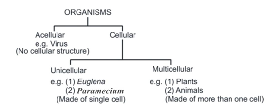
On the basis of number of cells living organisms can be divided into:
On the basis of complexity of organization, cells can be divided as Prokaryotes and Eukaryotes.
(i) Prokaryotic cells: These are primitive and incomplete cells. They have less developed nucleus without nuclear membrane & nucleolus. e.g. Bacteria.
(ii) Eukaryotic cells: These are well developed cells. They have advanced nucleus with nuclear membrane and nucleolus. e.g. Plants & animals.
Difference between Prokaryotic cells and Eukaryotic cells
| S.No. | Prokaryotic Cell | Eukaryotic Cell |
|---|---|---|
| 1. | Size of cell is generally small (1-10 μm). | Size of cell is generally large (50 to 100 μm). |
| 2. | Nuclear region is poorly defined, not surrounded by nuclear membrane and therefore called nucleoid and not nucleus. | Nuclear region is well defined, surrounded by a nuclear membrane. Therefore complete nucleus is present. |
| 3. | Contains single chromosome. | Contains more than one chromosome. |
| 4. | Nucleolus is absent. | Nucleolus is present. |
| 5. | Membrane bound cell organelles absent. | Membrane bound cell organelles such as mitochondria, plastids, endoplasmic reticulum, golgi apparatus, lysosomes, peroxisomes etc. are present. |
| 6. | Cell division takes place by fission or budding (no mitosis). | Cell division occurs by mitosis or meiosis. |
| 7. | Centrioles absent. | Centrioles present in animal cells. |
| 8. | Prokaryotic cells are found in bacteria and blue-green algae. | Eukaryotic cells are found in fungi, plant and animal cells. |
Shape, Size, Number, Volume Of Cell
The size, shape, number and volume of the cell is different among unicellular and multicellular organisms.
Cell Shape
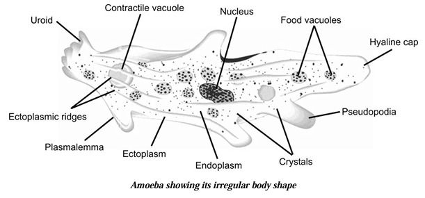
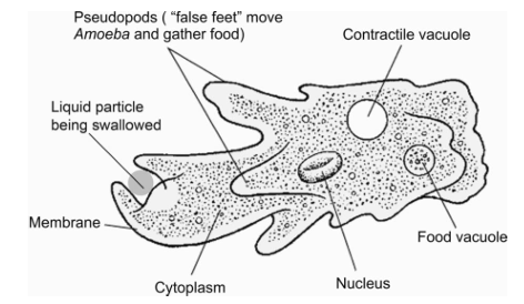
(a) Cell shape can be variable (e.g. Amoeba) or fixed (Euglena and Paramecium).
(b) In multicellular organisms, the shape depends on functional adaptations and partly on the surface tension, viscosity of protoplasm, the mechanical action exerted by adjoining cells and rigidity of cell membrane.
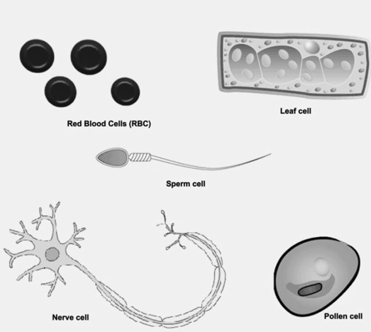
(c) The various shapes are:
| S.No. | Shape | Example |
|---|---|---|
| 1. | Spherical | Eggs of many animals |
| 2. | Spindle shaped | Smooth muscle fibre |
| 3. | Elongated | Nerve cells |
| 4. | Branched | Chromatophores or pigment cells of skin |
| 5. | Discoidal | Erythrocytes or RBC |
| 6. | Polyhedral | with 8, 12 or 14 sides |
Cell Size
Cell size vary. Some plant and animal cells are visible to the naked eyes but mostly seen with microscope since they are only few micrometers in diameter. The cells can be as small as 0.2 to 5 μm e.g. bacteria.
| Category | Example |
|---|---|
| Largest Cell | Ostrich Egg |
| Smallest Cell | Mycoplasma gallisepticum |
| Longest Cell | Nerve cell |
| Largest cell (plant) | Ovule of Cycas |
Unit of Measurement used in cell Biology
- 1 mm = 10-3 metre
- μm = micrometer
- 1 μm = 10-3 mm
- nm = nanometer
Cell Volume
The volume of a cell:
- (a) Is fairly constant for a particular cell type.
- (b) Is independent of the size of an organism.
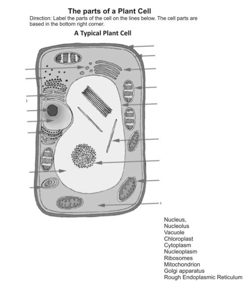
Difference Between Animal And Plant Cell
| S.No. | Animal Cell | Plant Cell |
|---|---|---|
| 1. | Animal cells are generally smaller in size. | Plant cells are larger than animal cells. |
| 2. | Cell wall is absent. | The plasma membrane of plant cells is surrounded by a rigid cell wall of cellulose and hemicellulose. |
| 3. | Except the protozoan (Euglena) no animal cell possesses plastids. | Plastids (Chloroplasts, Chromoplasts and Leucoplasts) are present. |
| 4. | Animal cells have a single highly complex and prominent Golgi apparatus. | Plant cells have many simpler units called dictyosomes. |
| 5. | Animal cells have centrosome and centrioles. | Plant cells lack centrosome and centrioles. |
| 6. | Vacuoles are smaller but more in number. | Vacuoles are larger but less in number (1-3) |
| 7. | Nucleus is mostly in the centre. | Nucleus is mostly towards the periphery. |
Components Of Cell
There is an occurrence of division of labour within a cell as they all got certain specific components called "Cell organelles" each of them perform a specific function.
There is division of labour present in multicellular organisms.
- This means that different types of cells present in the body of the organism carry out different functions.
- For Example: lungs perform the function of exchange of gases, stomach is for digestion of food.
- Division of labour is also seen within a cell.
- Specific structures that is cell organelles present inside the cell perform specific functions.
- For Example: mitochondria synthesize ATP or ribosome carry out protein synthesis.
- Interestingly, different types of cells have same organelles though they may differ in their functions.
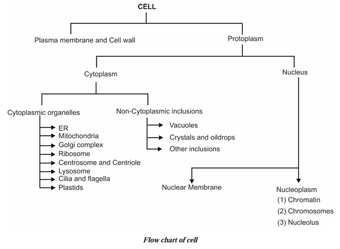
- The three basic components of all the cells are:
- Plasma Membrane
- Nucleus
- Cytoplasm
Detailed Structure Of Cell
Cell Membrane (plasma Membrane)
- Cell membrane is also called as plasma Membrane or Plasma lemma.
- It is the limiting boundary of each cell which separates the cytoplasm from its surroundings.
- It is found in both plant as well as animal cells.
- It is the outer most covering of a cell in case of animals and lies below the cell wall in case of plants.
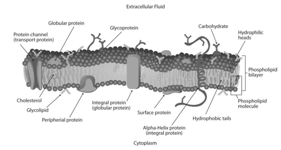
- It is made up of proteins and lipids where proteins are sandwiched between bilayer of lipids.
- Plasma membrane name was given by Nageli.
- Plasma membrane is selectively permeable in nature. It allows or permits the entry and exit of some materials in and out of the cell.
- Singer and Nicholson gave the fluid mosaic model of plasma membrane according to him it consists of a protein layer sandwiched between two layers of lipids. It is in quasifluid state. It is 75Å thick.
- It is flexible and can be folded, broken and reunited.
(i) Functions of plasma membrane:
(A) It regulates the movement of molecules inside and outside the cell.
(B) It helps in maintaining the distinct composition of the cell.
(ii) Transportation of molecules across the plasma membrane:
This can be done by following ways:
(A) Diffusion:
Movement of solutes or ions from higher concentration to lower concentration is called as diffusion. It does not require energy therefore it is called as passive transport.
(B) Osmosis:
The movement of solvent or water from higher concentration (solvent) to lower concentration (solvent) through a semipermeable membrane is called as osmosis or the movement of solvent or water from lower concentration to higher concentration of solution through a semipermeable membrane is called as osmosis. Osmosis can also be called as "diffusion of solvents".
- Endosmosis: Movement of solvent into the cell is called as endosmosis.
- Exosmosis: Movement of solvent outside the cell is called as exosmosis.
(iii) Types of solution on the basis of concentration:
(a) Isotonic solution: When the concentration of the solution outside is equal to the concentration of cytoplasm of the cell it is called as isotonic solution.
(b) Hypertonic solution: When the concentration of the solution outside the cell is more than that of inside the cell. Due to this cell looses water and becomes plasmolysed.
(c) Hypotonic solution: When the concentration of the solution outside the cell is lesser than that of cytoplasm of cell. Due to this cell swells up and bursts.
(d) Active Transport: It is the movement of molecules or ions from their lower concentration to higher concentration across the plasma membrane, i.e., it occurs against the concentration gradient. It often results in the accumulation of substances within the cell in higher concentrations than the outside concentration. It requires the utilization of energy in the form of ATP and also some membrane proteins acting as carrier molecules within the plasma membrane.
(a) Advantages of active transport:
- It helps the cell to absorb many selective ions or molecules inside it against concentration gradient.
- It is a very rapid process as compared to passive transport.
- It helps to maintain ionic and water balance between the cells and outside fluid.
- It helps to maintain action potential inside and outside the nerve membranes to enable the flow of stimulus across them.
(b) Types of Active Transport:
Active transport may occur by the following 2 modes:
- Endocytosis
- Exocytosis
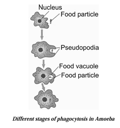
(i) Endocytosis: It is a process of intake of materials inside the cell through the plasma membrane. According to the nature of substances taken in, it is of following two types:
(a) Phagocytosis: It is the process by which the cell takes in solid food material through its plasma membrane. Therefore, the process is also called as cell-eating. All protozoans take in their food by this process.

(b) Pinocytosis: It is the process by which the cells takes in the fluid matter through its plasma membrane, therefore the process is called cell-drinking. In this process an invagination occurs at any point in the plasma membrane near the fluid source and small-pocket like structure is formed. The fluid is then engulfed inside the cell via this invagination and it forms a pinosome. The digestion of pinosome is then brought about by various enzymes of the cell that are present in the lysosomes.
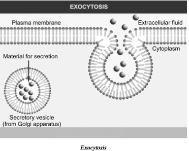
(ii) Exocytosis: It is the process of exudation or secretion of the waste materials out of the cell through plasma membrane. It is opposite of endocytosis. Exocytosis occurs in the cells of pancreas where the vacuoles containing enzymes move towards the membrane to discharge their contents to the exterior.
Plasmolysis
- When a plant cell is kept in hypertonic solution, cell loses water due to exosmosis.
- Shrinkage of cytoplasm and its contents occur.
- Cytoplasm gets detached from the cell wall and occupies one side of the cell.
- This phenomenon is called as plasmolysis.
Cell Wall
- It is the outermost covering of the plant cells.
- It is absent in animal cells.
- Cell wall is rigid, strong, thick, porous and non living structure. It is made up of cellulose and hemicellulose. Cell walls of two adjacent cells are joined by a layer called middle lamellae. It is made up of calcium and magnesium pectate.
- It has narrow pores, called pits, through which fine strands of cytoplasm, called plasmodesmata, are able to pass. These intercellular connections allow exchange of materials between living cell contents.
Functions of cell wall
- It provides definite shape to the cell.
- It provides strength to the cell.
- It is permeable and allows entry of molecules of different sizes.
- It is antigen specific.
- It has the characteristics of repair and regeneration.
Difference between cell wall and plasma membrane
| S.No. | Cell Wall | Plasma Membrane |
|---|---|---|
| 1. | It is present in plant cells only. | It is present in both animal and plant cells. |
| 2. | It is the outermost covering of plant cells. | It is the outermost covering of the animal cells. |
| 3. | It is present outside the plasma membrane. | It is present outside the cytoplasm. |
| 4. | Cell wall is rigid and comparatively thick. | It is comparatively flexible and thin. |
| 5. | It is made up of cellulose. | It is made up of lipids and proteins. |
| 6. | It is non-living and fully permeable. | It is living and selectively permeable. |
Protoplasm
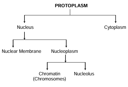
- It is a colourless, transparent, viscous, base liquid in which all cell organelles are suspended. Every cell is made up of protoplasm.
- The chemical structure of protoplasm was discovered in 1880 by Huxley in 1898 discovered that it is the physical basis of life.
- Protoplasm consists of:
- Nucleus
- Cytoplasm
Nucleus
- Nucleus is the most important cell organelle which directs and controls all its cellular activities.
- It is called as "Headquarter of the cell".
- It was discovered by "Robert Brown in 1831".
- In eukaryotes a well defined nucleus is present while in prokaryotes a well defined nucleus is absent.
- Prokaryotes contain a primitive nucleus.
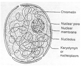
- It has double layered covering called as nuclear membrane.
- Nuclear membrane has pores which regulate the movement of materials in & out of the cell.
- Besides nuclear membrane nucleus also contains nucleolus and chromatin material and the substance filled inside the nucleus is nucleolus or karyolymph.
- Chromosomes or chromatin material consists of DNA which stores and transmits hereditary information for the cell to function, grow and reproduce.
Functions of the nucleus
- It controls all the metabolic activities of the cell and regulates the cell cycle.
- It helps in transmission of hereditary characters from parents to off springs.
- Nuclear membrane regulates the exchange of material between nucleoplasm and cytoplasm.
- DNA directs and control all the metabolic activities, therefore nucleus is called as brain of cell.
- Genes are the segment of DNA which are functional units of chromosome and act as unit of heredity (inheritance of characters).
- DNA control the synthesis of structure and enzymatic proteins. Changes in DNA (called gene mutations) produce variations.
Cytoplasm
- Cytoplasm was discovered by Kolliker in 1862.
- It is the site of both biosynthetic and catabolic pathways.
- It can be divided into two parts:
- Cytosol: Aqueous soluble part contains various fibrous proteins forming cytoskeleton.
- Cell organelles: Living part of the cells having definite shape, structure and function bounded by Plasma membrane.
Endoplasmic Reticulum (ER)
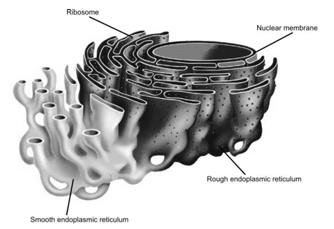
Nature and occurrence
- It is the network of membranes present in the cytoplasm.
- It was discovered by Porter, Claude and Fullam.
- These are present in all cells except prokaryotes and mammalian erythrocytes.
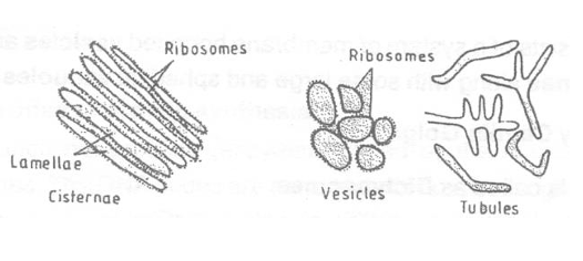
- They are made up of three components:
- Cisternae: These are long, flattened, parallely arranged, unbranched tubules. These form successive layers of nucleus. These are found in cells which are active in protein synthesis and are 40-50 µm in diameter.
- Vesicles: These are around or spherical they are found in synthetically active cells.
- Tubules
(a) Types:
Endoplasmic reticulum is of two types:
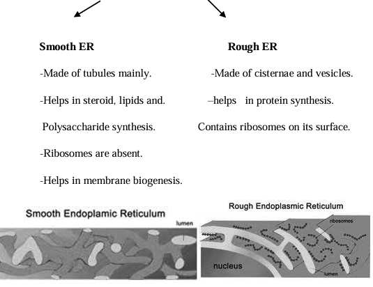
(b) Function of ER:
- It is the only organelle which can move within a cell so it serves as a channel for the transport of materials between various regions of cytoplasm and between cytoplasm and nucleus.
- It also function as a cytoplasmic framework to provide space for some of the biochemical activities. It forms endoskeleton of cell.
- It helps in synthesis of fats, steroids, cholesterol etc.
- It contains secretory proteins.
- SER plays a crucial role in detoxification of drugs and poisonous by-products.
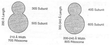
Ribosomes
Nature and occurrence:
- Discovered by Palade hence called Palade granules.
- Smallest cell organelle (230 Å in diameter).
- They are dense, spherical, granular particles occuring freely in cytosol (matrix) remain attached to ER.
- The main constituents of it are RNA and proteins.
- Two types of ribosomes are found 70S and 80S. 70S is found in prokaryotes which has two subunits (50S and 30S). 80S is found in eukaryotes which has two subunits (60S and 40S). Both the subunits of the ribosome remain attached when Mg2+ concentration increases but are separated when concentration decreases.
- All structural and functional proteins (enzymes) coded by the nuclear DNA, are synthesized upon cytoplasmic ribosomes. The DNA codes are transcripted into messenger RNA (mRNA) molecules in the chromosomes of the nucleus. mRNA molecules diffuse out into the cytoplasm and each becomes attached to several ribosomes which thus form a group called polyribosome or polyribosomes.
In this way each mRNA molecule brings about polymerization of specific protein molecules, with the help of ribosomes from amino acid molecules found in the Cytosol.
Function:
Ribosomes are site of protein synthesis and hence are called protein factories or engine of cells.
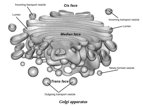
Golgi Apparatus
(Golgi body / Golgi Complex / Golgisome / Lipochondria)
Nature and Occurrence
- First discovered by Camello Golgi described by the name internal reticular apparatus.
- The apparatus lies near the nucleus. It is found in all eukaryotic cells except mammalian RBC's, bacteria, blue-green algae and mature sperms.
- In plants Golgi complex are known as dictyosomes.
- They are composed of:
- Cisternae: They are flattened, plate like tubules.
- Vesicles: They are spherical tubules
- Vacuoles: Large spherical peripherally occurring vesicles.
- Golgi vacuoles are constantly and rapidly renewed.
- Composed of lipids and fats mainly and that is why called lipochondria.
Functions:
- The main function of Golgi apparatus is secretory.
- It packages materials synthesized in the cell and dispatches from cell across the plasma membrane.
- It produces vacuoles or secretory vesicles which contain cellular secretions e.g. proteins, cellulose, melanin pigment, lactoprotein of milk, enzymes etc.
- It is also involved in the synthesis of cell wall, plasma membrane and lysosomes.
- Responsible for the development and growth of the reproductive cells in mammals.
- Anterior part of sperm i.e. acrosome is formed by them.
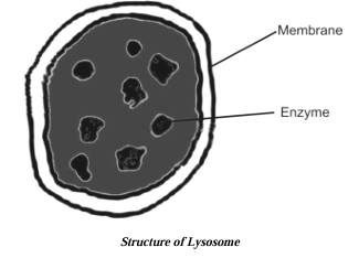
Lysosomes (enzymes Packets)
Nature and occurrence
- Discovered by Christian de Duve (1955) in rat liver cells.
- Are tiny spherical sac-like structures surrounded by a single, thin membrane of lipoproteins which contain digestive enzymes for intracellular digestion and waste disposal.
- Lysosomal enzymes are made by RER.
- It is abundant in digestive glands.
- The most important enzyme in lysosome is acid phosphatase.
Functions
- They serve as intracellular digestive system, hence, called digestive bags.
- Destroy any foreign material in the cell.
- Digestion of food, foreign material etc. by lysosome is called heterophagy.
- Remove the worn out and poorly working cell organelles by digesting them, removing cell debris and are hence known as demolition squad, scavengers, cellular housekeepers. Thus, forming a garbage disposal system of the cell.
- During breakdown of cell structure, when the cell gets damaged, lysosomes may burst and eat up their own cells. Therefore also called suicidal bags of cell.
Mitochondria
Nature and occurrence
- 'Mitochondrion' was first coined by Benda.
- Are tiny bodies of varying shapes and size (0.2 μm to 2 μm).
- Each mitochondria is bound by a double membrane.
- Outer membrane is porous, the inner membrane is thrown into folds called cristae and are studded with small round bodies known as F1 particles or oxysomes.
- The interior is filled with gel like matrix. Matrix contains small sized ribosomes, circular DNA molecule and phosphate granule.
- Mitochondria are absent in bacteria, RBC's of mammals.
- Oxysomes – generate ATP by oxidative phosphorylation.
Functions
- Are sites of cellular respiration.
- They synthesize energy-rich compounds (ATP) hence called 'power house of cell'.
- ATP is known as energy-carrier or energy-currency of the cell. It is cellular-fuel.
- ATP is used for the synthesis of chemical compounds e.g. (DNA replication, transcription, protein, carbohydrates and lipid synthesis) mechanical work (contraction of muscles, movement of cilia and flagella).
- Mitochondria makes some of their proteins therefore it is a semiautonomous organelle.

Plastid
Nature and occurrence
- It is double membranous discoidal structure, found only in plant cells.
- Term plastid was given by Haeckel.
- Chloroplast was discovered by A.V. Leeuwenhoek and named by Schimper.
- Besides being discoidal of rhombic in plant cells they occur in variable shapes like in algae they can be 'U' shaped, spiral, coiled, ribbon shaped etc.
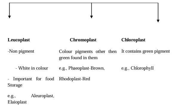
Depending upon the type of pigment present in them they are of following three types:
Chloroplast have Following Two Parts:
(i) Grana: It constitutes the lamellar system. These are found layered on top of each other, these stacks are called as Grana. Each granum of the chloroplast is formed by superimposed closed compartments called thylakoids.
Function: They are the sites of light reaction of photosynthesis as they contain photosynthetic pigment chlorophyll. In each thylakoid Quantasomes are present which are called as Photosynthetic units. Each quantasome possesses 230 chlorophyll molecules.
(ii) Stroma: It is a granular transparent substance also called as matrix. Grana are embedded in it. Besides Grana they also contain lipid droplets, starch grains, ribosomes etc.
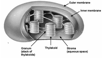
Function: This is the site of dark reaction of photosynthesis. Also helps in protein synthesis due to presence of ribosomes.
Cell Inclusions
These are non-living structures of the cytoplasm. These include reserve food in the form of grains (e.g. starch grains of potato tuber cells) or granules (e.g. glycogen in liver and muscle cells); or droplets (e.g. oil droplets in fat cells).
Vacuoles
- These are small sized but many in number in animal cells while plant cells have single large central-vacuole (which occupies 50-90% of the cell volume), so that the nucleus and cell organelles are pushed near the cell wall.
- A vacuole is formed of outer limiting membrane, called tonoplast.
Types of Vacuoles
Vacuoles are of two types viz. food vacuoles and contractile vacuole.
(i) Food vacuoles:: In Amoeba and amoeboid cells of higher animals the sacs containing ingested food particles fuse with lysosomes to form food vacuoles.
(ii) Contractile vacuole: Occur in some unicellular fresh water organisms e.g. Amoeba, Paramecium. They perform the function of osmoregulation.
Functions of Vacuole
- In animal cells they store water, glycogen and proteins.
- In unicellular animals like Amoeba and Paramecium they contain food material and maintain osmotic balance.
- Plant vacuoles are filled with cell sap containing sugars, amino acids, some proteins, minerals and metabolic wastes.
- In plant cells they provide turgidity and rigidity.
Peroxisomes
Nature and occurrence
- They are small (0.3 to 1.5 μm in diameter) and spherical organelles containing powerful oxidative enzymes.
- They are found in photosynthetic cell of plants, liver and kidney.
- They are bound by single membrane. There is a crystalline core in the centre of peroxisomes.
- The crystalline core is a crystallized protein called catalase.
Functions
- They detoxify or remove toxic substances from cell.
- In plant cells, they help in photorespiration.
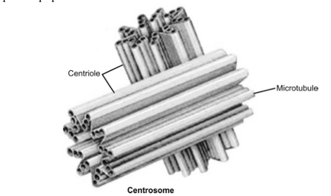
Centrosome
Nature and occurrence
- Found only in animal cell.
- It consists of two granules called centrioles.
- Centrioles are hollow, cylindrical structures which are made up of microtubules.
- In plant cells, polar caps perform the function of centrioles.
Functions
- Centrosome helps in cell division in animal cells as they are involved in the formation of spindle fibers.
- The microtubules of cilia and flagella originate and are borne by basal bodies or kinetosome formed by them.
- It develops the axial fibers of sperms.