In the Tissues chapter, Class 9 Science students explore how groups of cells form tissues in plants and animals. NCERT solutions for Class 9 Science make it easy to learn the differences between meristematic and permanent tissues. Our class 9 notes explain plant and animal tissues using detailed diagrams. Through class 9 science tuition, tutors simplify complex biological terms and structures. Interactive Class 9 tuitions focus on visual learning and concept retention through activities and quizzes. This topic strengthens understanding of human and plant anatomy, which is crucial for advanced biology learning.
Tissues
The higher plants and animals have highly complex bodies made up of various kinds of cells. These groups or clusters of cells are called tissues. Tissues improve the efficiency with which the body functions by allowing division of labour, that is, sharing of tasks. Each tissue is specialized for a particular job.
Thus, a tissue can be defined as a group of similar or dissimilar cells of common origin that perform or help to perform a common function.
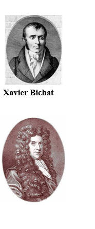
The term ‘tissue; was coined by N.Grew and was referred to animal tissue too by a French biologist Bichat. The study of tissue is called histology.
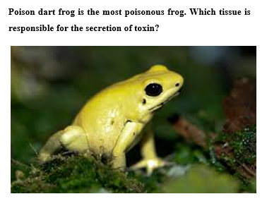
Poison dart frog is the most poisonous frog. Which tissue is responsible for the secretion of toxin?
Tissues
- The study of tissues is known as histology.
- The concept histology includes understanding of the structure of cells, tissues, organs and organ system, which can better by described as Microscopic Anatomy.
- Marie Xavier Bichat is known as the father of modern histology and pathology.
- He worked without a microscope and first introduced the notion of tissues.
- He maintained that diseases attacked tissues rather than organs.
Xavier Bichat
Tissue
Nehemiah Grew: (1641-1712) was an English plant anatomist and physiologist. His great work the anatomy of plants appeared in 1862. It was divided into four books: Anatomy of vegetable, anatomy of roots, Anatomy of trunks, and Anatomy of leaves, flowers, Fruits and Seeds. Anatomy of plants is especially notable for its descriptions of plant structure. He described nearly all key differences of the morphology of stem and root, showed that the flowers of Asteraceae are built of multiple units, and correctly hypothesissed that statement are male organs. Anatomy of plants also contains the first known microscopic description of pollens. Linnaeus named a genus of trees Grewia in his honour. He is called the father of plant anatomy.
- In multicellular organisms there is division of labor.
- A group of cells that are specialized to carry out a functions effectively.
- Tissue is a group of cells similar in structure and function. Various tissues of an organism work in co-ordination with each other in order to perform various functions.
- In plants and animals tissue are found but these tissues have differences on various aspects which are following:
Table: Differences between Plant and Animals Tissues
| S.No. | Plants Tissues | Animal Tissues |
| 1. | Dead supportive tissues are more abundant as compared to living tissues. | Living tissues are more common as compared to dead tissues. |
| 2. | They require less maintenance energy. | Animal require more maintenance energy. |
| 3. | Organisation is simple. | Organisation is complex with the development of more specialized and localized organs and organ systems. |
| 4. | Tissue organizations is towards stationary habit. | Tissue organization is towards high mobility. |
| 5. | There is differentiation of meristematic and permanent tissues. | Such differentiation is absent. |
Something Special
All the tissues of a plant which perform some general function irrespective of its position in plant body are considered to form
together a 'tissue system'.
According to Sacbs, there are three main plant tissues system :
(1) Epidermal Tissue System : It forms the outer covering of the plant. It performs manyfold function e.g., protection, absorption, excretion, secretion, gaseous exchange and control of transpiration etc.
(2) Ground Tissue System : The tissue that do not come under epidermal tissue system or vascular tissue system arc regarded to constitute the fundamental or the ground tissue system. In dicots, it consists hypodermis, cortex, endodermis, pericycle and pith.
(3) Vascular Tissue System : The central cylinder of the shoot or root surrounded by cortex is called stele. The varying number of vascular bundles formed inside the stele constitute vascular tissue system. It consists Xylem, phloem or bast and cambium.
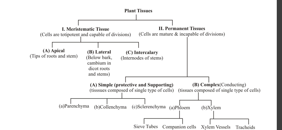
Plant Tissues
Mainly they are of two types:
(i) Meristematic (ii) Permanent
Plant Tissues
- Unlike animals, in plants growth is not uniform.
- Plants have demarcated regions of dividing and non dividing cells.
- Based on the dividing capacity of the tissue, the plant tissues can be broadly classified into:
- Meristematic Tissue
- Permanent Tissues
Meristematic Tissues (Meristems)
- The growth of the plant is due to dividing tissue, known as meristematic tissue.
- Depending on the region, where they are present, the meristematic tissue is classified as
- Apical
- Inter calary
- Lateral
Apical Meristem
- Apical meristem is present at the growing tips of stem and root.
- It brings about elongation of root and stem.
- It results in increase in the height of plants, which is called as primary growth.
- Apical meristem in shoot tip Apical Meristem in root tip
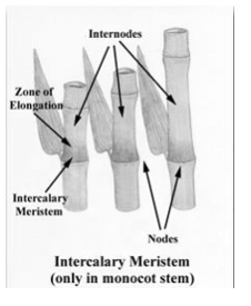
Intercalary Meristem
- Intercalary meristem is present at the base of the leaves or internodes.
- It results in the increase of length of the region.
Lateral Meristem
- Lateral meristem is present beneath the bark (cork cambium) and in the vascular bundles of dicot plants.
- It increases the diameter and girth of stem or root.
- This is called as secondary growth.
Permanent Tissue
- These tissues are derived from the meristematic tissue.
- When meristematic tissue loses its power of division and attains a definite form, it is called as permanent tissue.
- Permanent tissues are classified into two types:
- Simple permanent tissues which are made up of only one type of cells: Parenchyma, collenchyma and sclerenchyma.
- Complex permanent tissues which consists of more than one type of cells : Xylem and phloem.
Simple Permanent Tissue
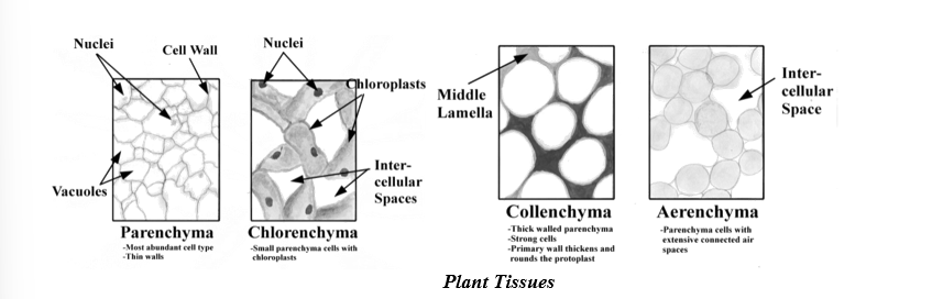
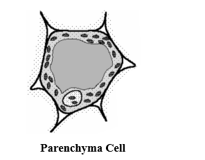
Parenchyma
- Parenchyma cells are living and form bulk of the plant body.
- They function in storage of food, photosynthesis and serves as packing tissue to fill the spaces between other tissues.
- Palisade parenchyma cells are elongated cells located mainly in leaves just below the epidermal tissue.
- Spongy mesophyll parenchyma cells occur below the one or two layers of palisade cells.
- Parenchyma Cell
- Ray parenchyma cells occur in wood rays, the structures that transport materials laterally within a woody stem.
- Parenchyma cells also occur within the xylem and phloem of vascular bundles.
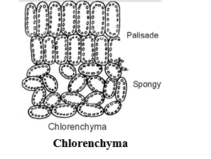
Chlorenchyma
- If chloroplast is present, the parenchyma tissue is called chlorenchyma.
- It performs photosynthesis, e.g.; the mesophyll cells of leaves.
Aerenchyma
- In aquatic plants large air cavities are present in the parenchyma and it is called as aerenchyma.
- It gives buoyancy to aquatic plants for floating in water, e.g., Lotus, Hydrilla.
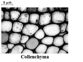
Collenchyma
- Consists of living cells.
- These cells are characterized by the deposition of extra cellulose at the corners of the cells.
- They are located in the petiole, mid ribs of dicot leaves and below the epidermis of dicot stems.
- It allows easy bending in various parts of the plant without actually breaking it.
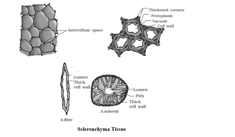
Sclerenchyma
- Sclerenchyma are dead cells and devoid of protoplasm.
- The walls of sclerenchyma cells are thickened due to deposition of lignin, such cell walls are called lignified.
- A conspicuous middle lamella is present between two sclerenchymatous cells. Middle lamella is a cementing substance containing pectin and lignin.
- Sclerenchyma cells are of two types; fibres and sclereids.
- Fibres are long narrow cells pointed at both ends and sclereids are irregular shaped.
- Most sclerenchyma cells are dead at maturity and therefore lack nuclei.
- They occur in the veins of leaves and in hard covering of seeds and nuts.
- Sclerenchyma provides mechanical strength to the plant.
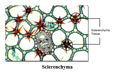
- They protect the plant from environmental forces like strong winds.
- The husk of coconut is made up of sclerenchyma tissues.
- Some sclerenchyma cells occur in the fruits of Pear. These cells (sclereids or stone cells) give pears their gritty texture.
Complex Permanent Tissue
Xylem
- It is a conducting tissue composed of four different type of cells :
- Tracheids
- Vessels
- Xylem parenchyma
- Xylem sclerenchyma (fibers).
- Except xylem parenchyma, all other xylem elements are dead with lignified cell walls.
- Xylem Components
- Tracheids are elongated cells with tapering ends. They conduct water from cell to cell via the pits present in their cell walls
Vessels are wider than tracheids.
- The transverse walls between the vessel elements are partially or completely dissolved to form a continuous pipe like structure.
- Xylem parenchyma stores food and helps in lateral conduction of water.
- The main function of xylem is to carry water and mineral salts dissolved in it, from roots to different parts of the plants.
- Lignified walls of xylem tissue give mechanical strength to the plant body.
Phloem
- Phloem cells conduct food from leaves to rest of the plant.
- Phloem cells are usually located outside the xylem.
- Phloem consists of four type of cells:
- Sieve tubes.
- Companion cells
- Phloem fibers
- Phloem Parenchyma
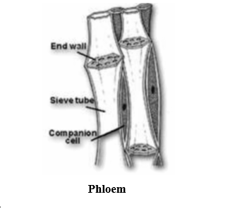
- Except for phloem fibers, all other phloem cells are living cells.
- Sieve tubes are long, thin walled cells placed end to end.
- Their end walls are perforated by many pores and are called sieve plates.
- The cytoplasm of one sieve tube is continuous with those of the sieve tube above and below through these pores.
- Companion cells are associated with the sieve tube and are small thin walled cells.
Phloem
Quick Digest
The sieve elements in angiosperms are sieve tubes arranged one above the other in distinct linear rows and walls and have sieve plates on their end walls and are associated with companion cells.
In gymnosperms and pteridophytes on their lateral walls, the companion cells are absent and there is no distinct arrangement of sieve to cells in linear rows-such sieve
They are connected with the sieve tube by numerous plasmodesmatas.
- Phloem transports food material synthesized due to photosynthesis in leaves, to different parts of the plant.
- Phloem fibres are sclerenchyma fibres. They provide mechanical strength.
| S.No. | Xylem | Phloem |
| 1. | Xylem parenchyma only living cells remaining cells are dead. | Phloem sclerenchyma only dead cells remaining all cells are living |
| 2. | It carries minerals, salts and water | An organic solution of sugars is translocated |
| 3. | The movement is only upward | The movement can be upward of downward. |
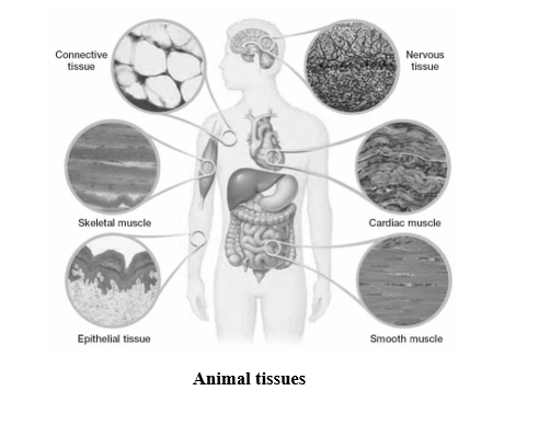
Animal Tissues
On the basis of function they perform in the body of multicellular animals, the animal tissues are classified as:
- Epithelial
- Connective
- Muscular
- Nervous
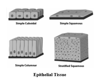
Epithelial Tissue
Lines, covers, and protects other tissues and organs.
- Cells are tightly packed together
- The presence of a non cellular basement membrane made of collagen.
- Simple epithelial tissue is composed of single layer of cells.
- Stratified epithelial tissue is made up of several layers of cells.
Functions of Epithelial Tissue
- Forms the outer layer of skin and protects the underlying cells.
- Forms lining of mouth, alimentary canal, respiratory organs, excretory organs and reproductive organs.
- Performs excretory and secretary function for example sweat, saliva, enzymes, hormones.
Epithelial tissues are classified as:
- Squamous epithelium
- Glandular epithelium
- Cuboidal epithelium
- Ciliated epithelium
- Columnar epithelium
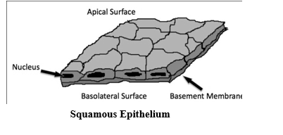
Squamous Epithelium
- Cells are very thin, flat and fit together like tiles.
- It forms lining of mouth, oesophagus, alveoli, blood vessels and lymphatic tubes, covering of skin.
- It protects the underlying parts of the body from mechanical injury or entry of germs.
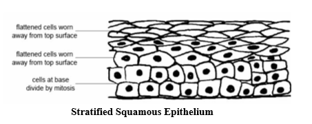
Stratified Squamous Epithelium
-
Cells are very thin, flat and fit together like tiles.
- It forms lining of mouth, oesophagus, alveoli, blood vessels and lymphatic tubes, covering of skin.
- It protects the underlying parts of the body from mechanical injury or entry of germs.
-
Found in skin and covers the external dry surface of the skin.
-
It protects the body from mechanical injury and prevents drying of underlying structures.
Cuboidal Epithelium
- Cells cube shaped – It occurs in
-
Kidney tubules
-
Ducts and secretory glands
- Germinal epithelium of ovary
- It helps in absorption, excretion and secretion and also provides mechanical support.
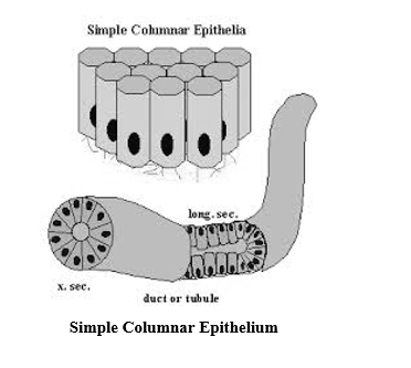
Columnar Epithelium
- Elongated cells, much longer than they are wide.
- The nuclei are present towards the base.
Simple Columnar Epithelium
- A single layer of cells that line the digestive tract, gallbladder, oviduct and secretory ducts of some glands.
- Found in organs where absorption and secretion occurs.
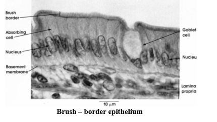
Brush – border epithelium
- Some times the free ends of cells have microvilli present, forming brush border.
- It facilitates the absorption of substances by increasing the surface area.
Glandular Epithelium
- Columnar epithelium is modified to secrete chemicals.
- Lining of salivary glands, pancreas, endocrine glands.
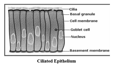
Ciliated Epithelium
- Certain columnar or cuboidal cells have cilia present on their free border, forming ciliated epithelium.
- It forms the lining of sperm duct, oviduct, trachea, bronchi, kidney tubules.
- It moves particles in one direction due to rhythmic beating of cilia in one direction.
Connective Tissue
- Connective tissue is binding and supporting tissue.
- Characterized by loosely spaced cells.
- Gel like intercellular substance called matrix forms the main bulk of connective tissue.
- Cell Matrix composed of two regions
- Ground that is liquid (sol), Gel, Gum or solid.
Loose Connective Tissue (Areolar)
- Gel like ground with both elastic (yellow) and non-elastic (white) fibres running through the matrix in many directions.
- It joins skin to muscles, fills spaces inside organs, found around muscles, blood vessels, nerves.
- It acts as packaging and supporting tissue between organs.
- It helps in repair of tissues after injury.
Dense Regular Connective Tissue
- Fibres are arranged orderly and densely in parallel rows.
- Occurs in tendons and ligaments
- Fibres mostly non-elastic.
- Tendons join skeletal muscles to bones.
- Ligaments join bones to bones
Adipose Tissue
- Formed by fat cells or adipocytes which are round cells containing large droplets of fat that almost fills the cell.
- Nucleus is pushed to one side of the cell.
- Occurs below the skin, between visceral organs and in yellow bone marrow.
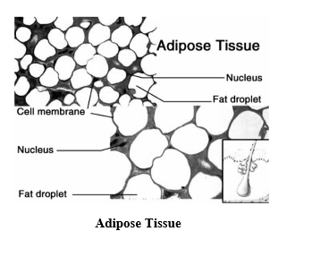
Functions of Adipose Tissue
- It serves as a fat reservoir.
- It keeps visceral organs in position and forms shock absorbing cushion around him.
- It regulates body temperature and act as an insulator.
- It provides shape to limbs and body.
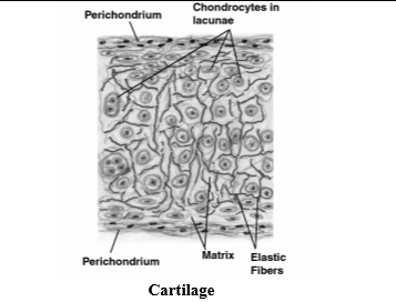
Cartilage
- It has matrix made up of proteins and calcium salts.
- Chondrocytes are found in fluid filled spaces called lacunae, within the matrix.
- Matrix have delicate network of fibres that may be elastic or non-elastic.
- Hyaline Cartilage – Example : on the ends of bones
- Elastic Cartilage – Example : ear pinna, rings of traches
- Non-elastic Cartilage– Example: nose cartilage, inter-vertebral discs.
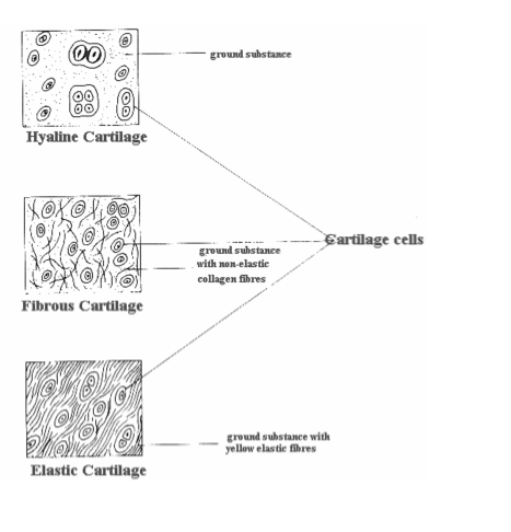
Functions of Cartilage
- Provides support and flexibility to the body parts.
- Prevents frictional wear and tear of bone tips to smoothen the surface of tips.
- Inter-vertebral discs made up of cartilage functions as cushions between the vertebra.
Bone
- It is porous, highly vascular, mineralized, hard and rigid tissue.
- Matrix of the bone is in the form of thin concentric rings called lamellae.
- Bone cells are called osteocytes or osteoblasts which are present between the lamellae.
- Matrix of bone is heavily coated with phosphate and carbonate salts of calcium and magnesium (hydroxyapatite).
- Each bone cell is enclosed in a small cavity called the lacuna.
- In mammals, the osteocytes are present in concentric rings around the central canal called the Haversian Canal.
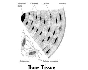
Functions of Bone
- Provides skeletal support to the body by forming endoskeleton.
- Protects vital organs like brain, heart, lungs.
- Serve as storage sites for calcium and phosphate.
Vascular Tissue (Blood)
- Blood is fluid connective tissue.
- Liquid matrix is plasma that contains
- 90% water.
- 10% plasma proteins such as albumin, globulin and fibrinogen, inorganic salts, organic substances, hormones, oxygen, glucose etc.
- Formed elements are blood corpuscles or cells.
- Erthrocytes or Red Blood Corpuscles – 48 billion (female) to 54 billion (male) cells/ml in the blood of humans.
- Mature Mammalian erythrocytes are enucleated.
- Presence of iron containing respiratory pigment haemoglobin.
- Plays an important role in transport of oxygen in the body.
- Leucocytes or White Blood Cells are of mainly two types: Agranuloytes and granulocytes
- Their number is 6000-9000 per/mm3.
- Agranulocytes.
- Lymphocytes and monocytes.
- Granulocytes.
- Neutrophils, basophils and eosinophils.
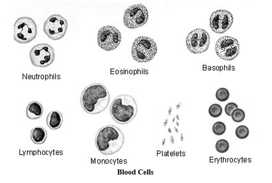
- They carry out function of body defense by engulfing bacteria and other foreign substances.
- Antibodies formation for immune response against pathogenic micro-organisms.
Platelets
- They are very small enucleated cells.
- Their number is 2,00,000 to 4,00,000 cells per/mm3.
- At the site of injury they help in clotting of blood.
Functions of Blood
- Transport nutrients, hormones and vitamins to the tissue and takes away excretory products from the tissues to liver and kidney.
- Conducts heat and regulates body temperature.
- RBCs carry oxygen to the tissues and bring back CO2 to the lungs.
- WBCs protect us from diseases by destroying harmful microorganisms entering our body.
- Blood platelets help in clotting of blood and prevents blood loss.
Lymph
- Colourless fluid containing WBCs in large number.
- After exchange of nutrients and gases, interstitial of fluid enters into lymph vessels and is called as lymph.
Functions of Lymph
- Lymph transports interstitial fluid from tissues to blood, which is rich in CO2 and wastes.
- Due to the presence of large numbers of lymphocytes, it protects the body against infection.
- Takes up proteins and fat droplets and transport them to the heart.
Serum
- It is a watery fluid which separates from blood when blood coagulates. It does not contain fibrinogen.
Muscle Tissue
- Forms the contractile tissue and made up of long muscle cells or fibers.
- Contractile protein is present in muscle cells which brings about contraction and relaxation of muscles.
- On the basis of their location, structure and function three types of muscle fibers are present :
- Striated or skeletal or voluntary muscles
- Smooth or unstriated or involuntary muscles
- Cardiac muscles

Striated Muscles
- Muscle fibers show alternate dark and light stripes thus termed as striated muscles.
- Attached to bones and are responsible for body movements.
- Muscles work according to our will therefore are also called voluntary muscles.
- Each striated muscle cell is multinucleated.
- Striated muscles provide locomotion and other voluntary movements of the body.
Smooth Muscles
- Spindle shaped, with a single nucleus present in the center of cell.
- Do not show any striations hence called smooth or unstriped muscles.
- Form the walls of visceral organs such as alimentary canal.
- Do not work according to our will so they are also called as involuntary muscles.
Cardiac Muscles
- Occur only in heart forming the walls of heart.
- Shows characteristics of both smooth and striated muscles.
- Muscle fibers are branched, striated and uninucleated.
- The space between cardiac muscles is filled with loose connective tissue and supplied with blood capillaries.
- Cardiac muscles contract and relax throughout life, pumping the blood to various parts of the body.
Nervous Tissue
- Made up of highly specialized cells called neurons.
- Neurons have the ability to receive stimuli and send the impulse to different parts of the body.
- Neuron has three parts.
- Cell body or cyton, Dendron and axons.
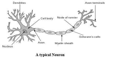
- Cell body contains nucleus and Nissl’s granules.
- Dendrons are short processes arising from the cyton. They carry impulse towards the cyton.
- Axon is a single long process and carry impulse away from cyton.
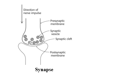
- There is a loose connection between the axon endings of one nerve cell and the dendrite of the next nerve cell, which is called as synapse.